WORD
document HERE
Within a decade Australians will be able to find out how good their genes are at fighting disease, which environmental risks they are susceptible to and steps they should take to prevent the onset of ill-health. And by the turn of the century it will be commonplace to have a bad combination of genes repaired to avoid disease.
'Then, Now…Imagine', a new report compiled by Research Australia in consultation with 10 of the country's leading health and medical researchers including two Nobel Prize winners and four Australians of the Year, predicts individual gene profiling from blood samples will revolutionise healthcare within ten years.
2006 Australian of the Year, Professor Ian Frazer, who discovered the technology that led to the newly released cervical cancer vaccine, said the upshot will be the ability to develop personalised healthcare plans – a roadmap for health from the day of birth.
"Doctors will be able to predict what health problems we might get so we can take appropriate precautions. They will also be able to assess what treatments will work best on an individual basis to achieve optimum health results. Long-term it will be possible to avoid certain diseases altogether through gene therapy," he said.
Sponsored by MBF, the report has been released by Research Australia to commemorate "Thank You" Day (14 November 2006), Australians' annual opportunity to send personal messages of appreciation to medical researchers whose work is special to them via www.thankyouday.org or 0428THANKS. "Thank You" Day is held each year with the support of the Macquarie Bank Foundation.
Therapeutic vaccines are also well advanced in development and involve re-educating the immune system to recognise cancer cells as intruders and attack them.
And over the next few decades we are likely see vaccines for many viral infections like HIV/AIDS and Hepatitis C, and for major diseases like diabetes. In fact Melbourne's Diabetes Vaccine Development Centre is about to start clinical trials for a new vaccine for Type 1 Diabetes.
This technology will also be applied to other disabilities. We will be able to reconnect electrical wiring in damaged spinal columns, stimulate nerve growth and allow messages to be relayed to the brain. Further into the future, this could ultimately allow quadriplegics and paraplegics to walk again. Other applications are likely to include correcting the faulty circuits that create epileptic episodes and creating transport systems for slow release of insulin to diabetics.
Professor Peter Doherty, Nobel Laureate 1996 – Discovered that T-cells, the foot soldiers in our bloodstream, were expert at killing cells that had viruses locked inside. This has led to new and better vaccines, healthy organ transplants and better treatment of conditions like Multiple Sclerosis and Diabetes.
Professor Ian Frazer, Australian of the Year 2006 – Gained international fame for developing the world's first vaccine to combat cervical cancer.
Dr Fiona Wood, Australian of the Year 2004 - Headed up the team of doctors who treated the burns victims of the 2002 Bali bombing. Her use of 'spray on skin' sped up the recovery process for those who had suffered horrific burns.
Professor Fiona Stanley, Australian of the Year 2003 - With Carol Bower, as part of an international collaboration, discovered the link between folate intake and spina bifida. This led to women being advised to increase folate intake before and during pregnancy and supplementation of some foods with folate.
Sir Gustav Nossal, Australian of the Year 2000 – Discovered the magic 'one cell-one antibody' rule which led to the development of effective new therapies for heart disease, breast cancer and severe arthritis.
Professor Terry Dwyer - Led the team which proved the link between a baby's sleeping position and Sudden Infant Death Syndrome. They found that a baby sleeping on its stomach has ten times the risk of SIDS than babies who sleep in other positions.
Professor Graeme Clark - Pioneered the multiple-channel cochlear implant which has brought hearing and speech understanding to tens of thousands of people with severe-to-profound hearing loss in more than 70 countries.
Professor Judith Whitworth – Discovered how steroids raise blood pressure. Former Chief Medical Officer for the Commonwealth Department of Health and Family Services and current Chair of the World Health Organisation (WHO) Global Advisory Committee on Health Research.
Professor John Shine – First to clone a human hormone gene and discovered a gene sequence, the Shine-Dalgarno sequence, which is important for the control of protein synthesis. Former Chair of the National Health and Medical Research Council and current member of the Prime Minister's Science, Engineering & Innovation Council.
Research Australia is a unique national alliance of more than 180 member and donor organisations with a common mission to make health and medical research a higher national priority. For more information on Research Australia visit www.researchaustralia.org.

"Plastic surgery patients are taking a proactive approach in making themselves happier by improving something that has truly bothered them," said Bruce Freedman, MD, ASPS Member Surgeon and study author. "While we are not saying that cosmetic plastic surgery alone is responsible for the drop in patients needing antidepressants, it surely is an important factor."
In the study, 362 patients had cosmetic plastic surgery – 17 percent or 61 patients were taking antidepressants. Six months after surgery, however, that number decreased 31 percent, down to 42 patients. In addition, 98 percent of patients said cosmetic plastic surgery had markedly improved their self-esteem.
All of the patients, who were primarily middle-aged women, had an invasive cosmetic plastic surgery procedure such as breast augmentation, tummy tuck or facelift. The authors did not identify any other major life changes that may have affected patients' use of antidepressants.
"We have just begun to uncover the various physical and psychological benefits of plastic surgery," said Dr. Freedman. "By helping our patients take control over something they were unhappy about, we helped remove a self-imposed barrier and ultimately improved their self-esteem."
The American Society of Plastic Surgeons is the largest organization of board-certified plastic surgeons in the world. With more than 6,000 members, the society is recognized as a leading authority and information source on cosmetic and reconstructive plastic surgery. ASPS comprises 94 percent of all board-certified plastic surgeons in the United States. Founded in 1931, the society represents physicians certified by The American Board of Plastic Surgery or The Royal College of Physicians and Surgeons of Canada.
Note: The study "Cosmetic Surgery and the Use of Antidepressant Medication" is being presented in electronic format, Sunday, Oct. 8 – Tuesday, Oct. 10, at the Moscone Convention Center, San Francisco. Reporters can register to attend Plastic Surgery 2006 and arrange interviews with presenters by logging on to www.plasticsurgery.org/news_room/Registration.cfm or by contacting ASPS Public Relations at (847) 228-9900 or in San Francisco, Oct. 7-11 at (415) 905-1730.
The new research comprehensively and convincingly makes the case that the small skull discovered in Flores, Indonesia, in 2003 does not represent a new species of hominid, as was claimed in a study published in Nature in 2004. Instead, the skull is most likely that of a small-bodied modern human who suffered from a genetic condition known as microcephaly, which is characterized by a small head.
"It's no accident that this supposedly new species of hominid was dubbed the 'Hobbit;'" said Robert R. Martin, PhD, Curator of Biological Anthropology at the Field Museum and lead author of the paper. "It is simply fanciful to imagine that this fossil represents anything other than a modern human." The new study is the most wide-ranging, multidisciplinary assessment of the problems associated with the interpretation of the 18,000-year-old Flores hominid yet to be published. The authors include experts on:
Significantly, the second most recent publication to conclude that the "Hobbit" was microcephalic--another multidimensional study that was published in the September 5, 2006, issue of Proceedings of the National Academy of Sciences--includes a co-author who was also a co-author of the original publication in Nature. That scientist, R.P. Soejono of the National Archaeological Research Center in Jakarta, Indonesia, now writes that the Flores hominid was microcephalic rather than a new hominid species.
Rewriting science
The starting point for the new research in Anatomical Record was the realization that the brain of the Flores skull (at 400 cc, the size of a grapefruit and less than one-third of the normal size for a modern human) is simply too small to fit anything previously known about human brain evolution. In addition, the stone tools found at the same site include types of tools that have only been reported for our own species, Homo sapiens.
Brain size of the Flores hominid, originally called Homo floresiensis, is known only from the main specimen discovered there, the LB1 skeleton. Skeletal fragments have been attributed to eight other individuals, but nothing can be said about their brain sizes. (They are small-bodied, but that has never been at issue.)
The new exhaustive research shows that the LB1 brain is simply too small to have been derived from H. erectus by evolutionary dwarfing, as was claimed by those wh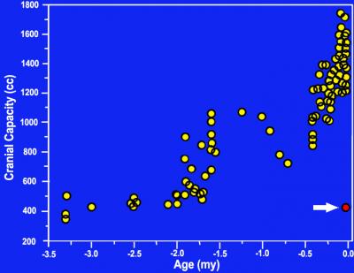 o
discovered it. In fact, the size
of the brain corresponds very closely to the average value for modern
human microcephalics. Microcephaly is a term that covers many
conditions. There are more than 400 different human genes for which
mutations can result in small brain size. Accordingly, there is a
correspondingly wide range of different syndromes that are recognized
in clinical practice. Many syndromes involve pronounced deficits
("low-functioning microcephaly"), but some have milder effects
("high-functioning microcephaly"), permitting survival into adulthood
and a surprising degree of behavioral competence in certain cases.
Microcephaly is often associated with severely reduced stature, but
some microcephalics have relatively normal body size.
o
discovered it. In fact, the size
of the brain corresponds very closely to the average value for modern
human microcephalics. Microcephaly is a term that covers many
conditions. There are more than 400 different human genes for which
mutations can result in small brain size. Accordingly, there is a
correspondingly wide range of different syndromes that are recognized
in clinical practice. Many syndromes involve pronounced deficits
("low-functioning microcephaly"), but some have milder effects
("high-functioning microcephaly"), permitting survival into adulthood
and a surprising degree of behavioral competence in certain cases.
Microcephaly is often associated with severely reduced stature, but
some microcephalics have relatively normal body size.
Because the LB1 skeleton is clearly that of an adult, it should obviously be compared with "high-functioning" modern human microcephalics rather than with "low-functioning" microcephalics who died early. The new study shows that skulls and brain casts from two modern human microcephalics who survived into adulthood are actually quite similar to those of the LB1 specimen. This supports the likelihood that LB1 was microcephalic.
Another area of controversy concerns the stone tools discovered in association with the Flores fossils. Initially, the discoverers claimed that the tools were sophisticated, as indeed they are. More recently, continuity has been claimed with tools from Mata Menge on Flores that are purportedly 800,000 years ago. This is simply implausible, according to the authors of the new research.
"Nobody has even claimed cultural continuity in stone tool technology over such a long period (800,000 to 18,000 years ago)," Dr. Phillips said. "To do so ignores the significance of tools found with the LB1 skeleton that were made with the advanced prepared-core technique, otherwise confined to Neanderthals and modern humans."
There has been too much media hype and not enough sound scientific evaluation surrounding this discovery, Dr. Martin concluded. "Science needs more balance and less acrimony as we continue to unravel this discovery."
Robert D. Marin, PhD, Curator of Biological Anthropology at The Field Museum, has devoted his career to studying primate development and evolution. In his quest to achieve a reliable reconstruction of primate evolutionary history, Dr. Martin has studied an extensive array of characteristics in the living species, including anatomical features, physiology, chromosomes and DNA. Dr. Martin has been particularly interested in the brain and reproductive biology, as these systems have been of special importance in primate evolution. With skeletal features, it is possible to include the fossil evidence and thus to include geological time in the picture. By studying living primates in the field in the forests of Africa, Madagascar, Brazil and Panama, Dr. Martin has also been able to include behavior and ecology in an overall synthesis. That synthesis was first presented in his textbook Primate Origins and Evolution, published by Princeton University Press in 1990. Since then, he has been working on refinements in several different directions. Photo by John Weinstein, Courtesy of The Field Museum (Negative # GN90075_36Ac)
• Jim Phillips
James Phillips, PhD, Departments of Anthropology at the University of Illinois at Chicago and the Field Museum, is an expert on stone tools. Initially, the discoverers of the Flores fossils claimed that the tools found in association with the fossils were sophisticated, as indeed they are. More recently, continuity has been claimed with tools from Mata Menge on Flores that are purportedly 800,000 years ago. This is simply implausible, according to the authors of the new research. "Nobody has even claimed cultural continuity in stone tool technology over such a long period (800,000 to 18,000 years ago)," Dr. Phillips said. "To do so ignores the significance of tools found with the LB1 skeleton that were made with the advanced prepared-core technique, otherwise confined to Neanderthals and modern humans."
"People often think breast reduction is an elective cosmetic procedure, but the majority of women seeking this surgery are legitimately debilitated by their breasts," said Michael Wheatley, MD, ASPS Member Surgeon and paper co-author. "The criteria most insurance companies use is not supported by medical literature and eliminates a large number of women from coverage, forcing them to fend for themselves."
Most insurance companies require patients to exhibit specific signs and symptoms prior to approving breast reduction as medically necessary. The amount of tissue removed to relieve symptoms associated with overly large breasts is the most controversial of all insurance criteria.
The authors reviewed the breast reduction policies of 87 health insurance companies. Despite contrary medical studies, 85 companies require a minimum amount of tissue to be removed to cover the procedure – 49 of these companies require a minimum amount to be removed independent of the patient's height and weight.
According to published studies, although most patients have a one-and-a-half to two cup size reduction, the amount of tissue removed, body weight, level of obesity, or bra cup size do not affect the benefits that patients receive from breast reduction.
Many insurance companies require that patients exhibit all of the following symptoms to receive coverage for breast reduction: back, neck, shoulder, and arm pain; rashes; bra strap grooves; and numbness in the upper torso. The authors found that while most patients suffer from many of these symptoms, rarely do they exhibit all.
According to Dr. Wheatley, most patients are women between 30 and 50 years old who have had upper skeletal pain for years. Many of them have tried various treatments, including physical therapy and pain medications, to manage the pain before turning to breast reduction. However, many of these women are still turned away by their insurance companies.
According to ASPS statistics, more than 114,000 breast reductions were performed in 2005.
The American Society of Plastic Surgeons is the largest organization of board-certified plastic surgeons in the world. With more than 6,000 members, the society is recognized as a leading authority and information source on cosmetic and reconstructive plastic surgery. ASPS comprises 94 percent of all board-certified plastic surgeons in the United States. Founded in 1931, the society represents physicians certified by The American Board of Plastic Surgery or The Royal College of Physicians and Surgeons of Canada.
Note: The study "Reduction Mammoplasty: A Review of Managed Care Medical Policy Criteria" is being presented Sunday, Oct. 8, 3:04 p.m., at the Moscone Convention Center, San Francisco. Reporters can register to attend Plastic Surgery 2006 and arrange interviews with presenters by logging on to www.plasticsurgery.org/news_room/Registration.cfm or by contacting ASPS Public Relations at (847) 228-9900 or in San Francisco, Oct. 7-11 at (415) 905-1730. VIRGINIA BEACH, VA (October 9, 2006) -- The long-lived naked mole-rat shows much higher levels of oxidative stress and damage and less robust repair mechanisms than the short-lived mouse, findings that could change the oxidative stress theory of aging.
The new study comparing the naked mole-rat, which has a life span of 28 years, and the mouse, which has a lifespan of three years, will be presented Oct. 8 at The American Physiological Society conference, Comparative Physiology 2006: Integrating Diversity. The results fly in the face of the oxidative stress theory of aging, which holds that damage caused by oxidative stress is a significant contributor to the aging process.
Under this theory, naked mole-rats should be better at preventing or repairing oxidative stress than their much shorter-lived cousin, the mouse. The study, "High oxidative damage levels in the longest-living rodent, the naked mole-rat," was done by Blazej Andziak and Rochelle Buffenstein, of The City College of New York, Timothy P. O'Connor, of Weill Medical College of Cornell University, and Asish P. Chaudhiri and Holly Van Remmen of the University of Texas Health Science Center, San Antonio. The study was presented during a poster session on October 8.
Don't toss the oxidative stress theory of aging out the window just yet, but prepare to modify it, said Buffenstein, the senior author. Her team suspects that the naked mole-rat's longevity stems from its ability to defend against acute bouts of oxidative stress. That is, the kind of oxidation that happens because of an unusual occurrence rather than the kind that happens as a result of normal aerobic respiration.
For example, when hydrogen peroxide is added to a culture containing naked mole-rat fibroblast cells, they remain viable and appear to repair the acute damage more rapidly than shorter-lived animals, explained Buffenstein.
What is old age?
We know that all organisms age and die. It's such an inevitable course of events that most of us spend more time thinking about how to hide the wrinkles and gray hair than we do about what our cells are actually doing to usher us to the end. Physiologists are looking at molecules and cells to understand this process.
One way to look at aging is to compare closely related organisms with different life spans. That's why it made sense to compare mole-rats and mice: They're the same size and they're rodents, but the mole-rat lives to 28 years, about nine-times longer than the mouse.
"Mole-rats must have something happening at the biochemical level to allow them to do this," said Andziak, the study's lead author. Specifically, he wanted to see if oxidative stress could explain the difference.
Oxidative stress occurs during metabolism when oxygen (O2) splits into single oxygen atoms, known as free radicals. These oxygen atoms may circulate by themselves, or combine with other atoms and molecules to form reactive oxygen species (ROS). ROS can damage DNA, lipids and proteins thus impairing normal cellular function. Antioxidants help to neutralize ROS, thus restricting the potential of ROS to damage biological molecules.
Mole-rat has more oxidative stress
The study compared two-year-old naked mole-rats to four-month-old mice. The researchers chose those ages so that the animals would be equivalent ages relative to their maximum lifespans, Andziak said.
First, the researchers compared the ratio of reduced glutathione, an antioxidant, to oxidized glutathione. As the body uses up its reduced glutathione to fight oxidative stress, the pool of oxidized glutathione increases. This ratio of reduced to oxidized glutathione is thus an indicator of oxidative stress: the greater the ratio, the less oxidative stress has occurred. The oxidative stress theory predicts that in naked mole-rats this ratio will be higher than in mice.
When the researchers measured this ratio in the liver, they found that the opposite was true. Mole-rats had less reduced glutathione and thus a lower ratio, indicating the mole-rat experienced much more oxidative stress. These results fit with the findings of a previous study in which Andziak found that naked mole-rats did not have superior antioxidant capacity when compared to mice. Mole-rats had much lower activity of the ubiquitous antioxidant enzyme, cellular glutathione peroxidase.
Mole-rat shows greater oxidative damage
The researchers next looked at how much damage the oxidation had caused. It is possible, they reasoned, that the mole-rat suffers greater oxidative stress, but its physiology had somehow prevented damage from occurring.
The researchers measured oxidative damage in lipids, DNA and proteins and found that naked mole-rats showed much greater levels of damage to each of these biological molecules, in all tissues assayed, when compared to mice. The study found multiple signs of lipid damage: The level of isoprostanes found in the urine was 10 times higher in the naked mole-rat, the level of malondialdehyde in liver tissue was twice as high and isoprostane levels in heart tissue was two-and-a-half times the level of the mice.
The researchers found significantly more protein damage in the kidney and in the heart. DNA damage was greater in the kidney and liver.
"All of the classical measures of oxidative stress are higher in the mole-rat," Andziak concluded. "Given that naked mole-rats live an order of magnitude longer than predicted based on their body size, our findings strongly suggest that mechanisms other than attenuated oxidative stress may explain the impressive longevity of this species."
Next steps
The next step is to determine how the mole-rats manage to live with the damage caused by oxidative stress. Buffenstein said she suspects that the mole-rat is able to fend off the occasional oxidative insult that can occur, and that may be more important than what happens with the steady-state levels of oxidative stress that result from normal metabolic activity.
Buffenstein theorizes that the naked mole-rats in her laboratory suffer higher levels of oxidative stress than they would in their natural underground habitat, where they encounter much lower levels of oxygen. But this exposure at an early age may provide some protection against acute oxidative stress and may be of considerable importance in their resistance to bursts oxidative stressors throughout their lives, she said.
"The naked mole-rat, with its surprisingly long lifespan and remarkably delayed aging, seems like the perfect model to provide answers about how we age and how to retard the aging process," Buffenstein said. "This animal may one day provide the clues to how we can significantly extend life."
APS provides a wide range of research, educational and career support and programming to further the contributions of physiology to understanding the mechanisms of diseased and healthy states. In 2004, APS received the Presidential Award for Excellence in Science, Mathematics and Engineering Mentoring.
11:50 07 October 2006
Within a decade Australians will be able to find out how good their genes are at fighting disease, which environmental risks they are susceptible to and steps they should take to prevent the onset of ill-health. And by the turn of the century it will be commonplace to have a bad combination of genes repaired to avoid disease.
'Then, Now…Imagine', a new report compiled by Research Australia in consultation with 10 of the country's leading health and medical researchers including two Nobel Prize winners and four Australians of the Year, predicts individual gene profiling from blood samples will revolutionise healthcare within ten years.
2006 Australian of the Year, Professor Ian Frazer, who discovered the technology that led to the newly released cervical cancer vaccine, said the upshot will be the ability to develop personalised healthcare plans – a roadmap for health from the day of birth.
"Doctors will be able to predict what health problems we might get so we can take appropriate precautions. They will also be able to assess what treatments will work best on an individual basis to achieve optimum health results. Long-term it will be possible to avoid certain diseases altogether through gene therapy," he said.
Sponsored by MBF, the report has been released by Research Australia to commemorate "Thank You" Day (14 November 2006), Australians' annual opportunity to send personal messages of appreciation to medical researchers whose work is special to them via www.thankyouday.org or 0428THANKS. "Thank You" Day is held each year with the support of the Macquarie Bank Foundation.
Five other
key forecasts are:
- Growing new body parts
- Smart drugs
- A Raft of New Vaccines
Therapeutic vaccines are also well advanced in development and involve re-educating the immune system to recognise cancer cells as intruders and attack them.
And over the next few decades we are likely see vaccines for many viral infections like HIV/AIDS and Hepatitis C, and for major diseases like diabetes. In fact Melbourne's Diabetes Vaccine Development Centre is about to start clinical trials for a new vaccine for Type 1 Diabetes.
- Building New Nerve Pathways
This technology will also be applied to other disabilities. We will be able to reconnect electrical wiring in damaged spinal columns, stimulate nerve growth and allow messages to be relayed to the brain. Further into the future, this could ultimately allow quadriplegics and paraplegics to walk again. Other applications are likely to include correcting the faulty circuits that create epileptic episodes and creating transport systems for slow release of insulin to diabetics.
- Operating before birth
--------------------------------------
You can download the
full 'Then, Now…Imagine' Report from www.thankyouday.org.
From 9 October until 17 November you can also send you personal message
of thanks to Australia's health and medical researchers via the website
or you can text it to 0428THANKS.
Scientists Who
Contributed To 'Then, Now…Imagine'
Dr Robin Warren, Nobel Laureate 2005 - with Barry
Marshall,
proved a bacteria called helicobacter pylori caused gastritis and
stomach ulcers and that most ulcers could be permanently cured with
antibiotics. Professor Peter Doherty, Nobel Laureate 1996 – Discovered that T-cells, the foot soldiers in our bloodstream, were expert at killing cells that had viruses locked inside. This has led to new and better vaccines, healthy organ transplants and better treatment of conditions like Multiple Sclerosis and Diabetes.
Professor Ian Frazer, Australian of the Year 2006 – Gained international fame for developing the world's first vaccine to combat cervical cancer.
Dr Fiona Wood, Australian of the Year 2004 - Headed up the team of doctors who treated the burns victims of the 2002 Bali bombing. Her use of 'spray on skin' sped up the recovery process for those who had suffered horrific burns.
Professor Fiona Stanley, Australian of the Year 2003 - With Carol Bower, as part of an international collaboration, discovered the link between folate intake and spina bifida. This led to women being advised to increase folate intake before and during pregnancy and supplementation of some foods with folate.
Sir Gustav Nossal, Australian of the Year 2000 – Discovered the magic 'one cell-one antibody' rule which led to the development of effective new therapies for heart disease, breast cancer and severe arthritis.
Professor Terry Dwyer - Led the team which proved the link between a baby's sleeping position and Sudden Infant Death Syndrome. They found that a baby sleeping on its stomach has ten times the risk of SIDS than babies who sleep in other positions.
Professor Graeme Clark - Pioneered the multiple-channel cochlear implant which has brought hearing and speech understanding to tens of thousands of people with severe-to-profound hearing loss in more than 70 countries.
Professor Judith Whitworth – Discovered how steroids raise blood pressure. Former Chief Medical Officer for the Commonwealth Department of Health and Family Services and current Chair of the World Health Organisation (WHO) Global Advisory Committee on Health Research.
Professor John Shine – First to clone a human hormone gene and discovered a gene sequence, the Shine-Dalgarno sequence, which is important for the control of protein synthesis. Former Chair of the National Health and Medical Research Council and current member of the Prime Minister's Science, Engineering & Innovation Council.
Research Australia is a unique national alliance of more than 180 member and donor organisations with a common mission to make health and medical research a higher national priority. For more information on Research Australia visit www.researchaustralia.org.
Some
patients stop needing
antidepressant medication after having plastic surgery
Study presented at American Society of Plastic Surgeons annual meeting
SAN FRANCISCO – It has been proven that plastic surgery can
improve
self-esteem, but can it also act as a natural mood enhancer? A
significant number of patients stopped taking antidepressant medication
after undergoing plastic surgery, according to a study presented today
at the American Society of Plastic Surgeons (ASPS) Plastic Surgery 2006
conference in San Francisco.
Study presented at American Society of Plastic Surgeons annual meeting
"Plastic surgery patients are taking a proactive approach in making themselves happier by improving something that has truly bothered them," said Bruce Freedman, MD, ASPS Member Surgeon and study author. "While we are not saying that cosmetic plastic surgery alone is responsible for the drop in patients needing antidepressants, it surely is an important factor."
In the study, 362 patients had cosmetic plastic surgery – 17 percent or 61 patients were taking antidepressants. Six months after surgery, however, that number decreased 31 percent, down to 42 patients. In addition, 98 percent of patients said cosmetic plastic surgery had markedly improved their self-esteem.
All of the patients, who were primarily middle-aged women, had an invasive cosmetic plastic surgery procedure such as breast augmentation, tummy tuck or facelift. The authors did not identify any other major life changes that may have affected patients' use of antidepressants.
"We have just begun to uncover the various physical and psychological benefits of plastic surgery," said Dr. Freedman. "By helping our patients take control over something they were unhappy about, we helped remove a self-imposed barrier and ultimately improved their self-esteem."
-------------------------------
For referrals to ASPS Member Surgeons certified by the
American
Board of Plastic Surgery, call 888-4-PLASTIC (475-2784) or visit www.plasticsurgery.org
where you can also learn more about cosmetic and reconstructive plastic
surgery.
The American Society of Plastic Surgeons is the largest organization of board-certified plastic surgeons in the world. With more than 6,000 members, the society is recognized as a leading authority and information source on cosmetic and reconstructive plastic surgery. ASPS comprises 94 percent of all board-certified plastic surgeons in the United States. Founded in 1931, the society represents physicians certified by The American Board of Plastic Surgery or The Royal College of Physicians and Surgeons of Canada.
Note: The study "Cosmetic Surgery and the Use of Antidepressant Medication" is being presented in electronic format, Sunday, Oct. 8 – Tuesday, Oct. 10, at the Moscone Convention Center, San Francisco. Reporters can register to attend Plastic Surgery 2006 and arrange interviews with presenters by logging on to www.plasticsurgery.org/news_room/Registration.cfm or by contacting ASPS Public Relations at (847) 228-9900 or in San Francisco, Oct. 7-11 at (415) 905-1730.
Compelling
evidence demonstrates
that 'Hobbit' fossil does not represent a new species of hominid
Most complete, interdisciplinary study published on raging controversy about fossil found in Flores, Indonesia
CHICAGO --
What may well turn out to
be the definitive work in a debate that has been raging in
palaeoanthropology for two years will be published in the November 2006
issue of Anatomical Record. Most complete, interdisciplinary study published on raging controversy about fossil found in Flores, Indonesia
The new research comprehensively and convincingly makes the case that the small skull discovered in Flores, Indonesia, in 2003 does not represent a new species of hominid, as was claimed in a study published in Nature in 2004. Instead, the skull is most likely that of a small-bodied modern human who suffered from a genetic condition known as microcephaly, which is characterized by a small head.
"It's no accident that this supposedly new species of hominid was dubbed the 'Hobbit;'" said Robert R. Martin, PhD, Curator of Biological Anthropology at the Field Museum and lead author of the paper. "It is simply fanciful to imagine that this fossil represents anything other than a modern human." The new study is the most wide-ranging, multidisciplinary assessment of the problems associated with the interpretation of the 18,000-year-old Flores hominid yet to be published. The authors include experts on:
- scaling effects of body size, notably with respect to the brain: Dr. Martin and Ann M. MacLarnon, PhD, School of Human & Life Sciences, Roehampton University in London;
- clinical and genetic aspects of human microcephaly: William B. Dobyns, PhD, Department of Human Genetics, University of Chicago; and
- stone tools: James Phillips, PhD, Departments of Anthropology at the University of Illinois at Chicago and the Field Museum.
Significantly, the second most recent publication to conclude that the "Hobbit" was microcephalic--another multidimensional study that was published in the September 5, 2006, issue of Proceedings of the National Academy of Sciences--includes a co-author who was also a co-author of the original publication in Nature. That scientist, R.P. Soejono of the National Archaeological Research Center in Jakarta, Indonesia, now writes that the Flores hominid was microcephalic rather than a new hominid species.
 |
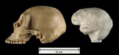 |
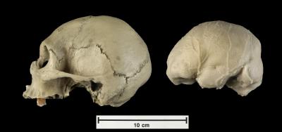
| Skull cast and cast of the endocranial cavity (endocast) from the Royal College of Surgeons in London of a modern adult human who suffered from microcephaly. It is strikingly similar to the following skull and endocast of a 32-year-old microcephalic woman. Together, the two specimens provide evidence that the LB1 skull from Flores, which is so similar to these two specimens, could also have been a microcephalic adult. Photo by John Weinstein, Courtesy of The Field Museum (Negative # Z94438_07Ad) | Skull cast and cast of the endocranial cavity (endocast) from a 32-year old woman who had the body size of a 12-year-old child. She lived in Lesotho, a county in Southern Africa, and these casts are part of The Field Museum's collection (Accession Nos. A219679 And A219680). This specimen and the one above have a relatively normal exterior appearance despite their very small size. Together they demonstrate that the LB1 skull from Flores could also have been an adult who suffered from microcephaly. Photo by John Weinstein, Courtesy of The Field Museum (Negative # Z94438_01Ad) |
The starting point for the new research in Anatomical Record was the realization that the brain of the Flores skull (at 400 cc, the size of a grapefruit and less than one-third of the normal size for a modern human) is simply too small to fit anything previously known about human brain evolution. In addition, the stone tools found at the same site include types of tools that have only been reported for our own species, Homo sapiens.
Brain size of the Flores hominid, originally called Homo floresiensis, is known only from the main specimen discovered there, the LB1 skeleton. Skeletal fragments have been attributed to eight other individuals, but nothing can be said about their brain sizes. (They are small-bodied, but that has never been at issue.)
The new exhaustive research shows that the LB1 brain is simply too small to have been derived from H. erectus by evolutionary dwarfing, as was claimed by those wh
o
discovered it. In fact, the size
of the brain corresponds very closely to the average value for modern
human microcephalics. Microcephaly is a term that covers many
conditions. There are more than 400 different human genes for which
mutations can result in small brain size. Accordingly, there is a
correspondingly wide range of different syndromes that are recognized
in clinical practice. Many syndromes involve pronounced deficits
("low-functioning microcephaly"), but some have milder effects
("high-functioning microcephaly"), permitting survival into adulthood
and a surprising degree of behavioral competence in certain cases.
Microcephaly is often associated with severely reduced stature, but
some microcephalics have relatively normal body size. Because the LB1 skeleton is clearly that of an adult, it should obviously be compared with "high-functioning" modern human microcephalics rather than with "low-functioning" microcephalics who died early. The new study shows that skulls and brain casts from two modern human microcephalics who survived into adulthood are actually quite similar to those of the LB1 specimen. This supports the likelihood that LB1 was microcephalic.
This
graph shows the cranial capacities in cubic centimeters for 118 fossil
hominids plotted against time, extending back almost 3.5 million years.
The arrow indicates the highly incongruous value reported in Nature by
Brown et al. in 2004 for H. floresiensis, described as an insular dwarf
derived from Homo erectus. The relatively tiny brain size only 18,000
years ago does not fit into known patterns of hominid brain size and
development. It is "off the chart." Graph by
Robert D. Martin, Courtesy of The Field Museum
Also, it has been claimed that LB1 had unusually large teeth
("megadonty"). However, it turns out that the teeth are not
particularly large, after allowing for the expected effect of dwarfing.
They are actually closely similar in size to those of a modern human
microcephalic. Another area of controversy concerns the stone tools discovered in association with the Flores fossils. Initially, the discoverers claimed that the tools were sophisticated, as indeed they are. More recently, continuity has been claimed with tools from Mata Menge on Flores that are purportedly 800,000 years ago. This is simply implausible, according to the authors of the new research.
"Nobody has even claimed cultural continuity in stone tool technology over such a long period (800,000 to 18,000 years ago)," Dr. Phillips said. "To do so ignores the significance of tools found with the LB1 skeleton that were made with the advanced prepared-core technique, otherwise confined to Neanderthals and modern humans."
There has been too much media hype and not enough sound scientific evaluation surrounding this discovery, Dr. Martin concluded. "Science needs more balance and less acrimony as we continue to unravel this discovery."
________________
•
Robert D. MartinRobert D. Marin, PhD, Curator of Biological Anthropology at The Field Museum, has devoted his career to studying primate development and evolution. In his quest to achieve a reliable reconstruction of primate evolutionary history, Dr. Martin has studied an extensive array of characteristics in the living species, including anatomical features, physiology, chromosomes and DNA. Dr. Martin has been particularly interested in the brain and reproductive biology, as these systems have been of special importance in primate evolution. With skeletal features, it is possible to include the fossil evidence and thus to include geological time in the picture. By studying living primates in the field in the forests of Africa, Madagascar, Brazil and Panama, Dr. Martin has also been able to include behavior and ecology in an overall synthesis. That synthesis was first presented in his textbook Primate Origins and Evolution, published by Princeton University Press in 1990. Since then, he has been working on refinements in several different directions. Photo by John Weinstein, Courtesy of The Field Museum (Negative # GN90075_36Ac)
• Jim Phillips
James Phillips, PhD, Departments of Anthropology at the University of Illinois at Chicago and the Field Museum, is an expert on stone tools. Initially, the discoverers of the Flores fossils claimed that the tools found in association with the fossils were sophisticated, as indeed they are. More recently, continuity has been claimed with tools from Mata Menge on Flores that are purportedly 800,000 years ago. This is simply implausible, according to the authors of the new research. "Nobody has even claimed cultural continuity in stone tool technology over such a long period (800,000 to 18,000 years ago)," Dr. Phillips said. "To do so ignores the significance of tools found with the LB1 skeleton that were made with the advanced prepared-core technique, otherwise confined to Neanderthals and modern humans."
Insurance
companies deny
medically necessary breast reductions based on random, unproven criteria
Study Presented at American Society of Plastic Surgeons annual Meeting
SAN FRANCISCO – What
if you couldn't perform daily activities, such
as exercising or running with your children, because of overly large
breasts that caused unending pain? Despite existing scientific studies
that outline the medical necessity for breast reduction, many insurance
companies are denying thousands of women the procedure each year
because of rigid, unfounded conditions to secure coverage, according to
a study presented today at the American Society of Plastic Surgeons
(ASPS) Plastic Surgery 2006 conference in San Francisco.
Study Presented at American Society of Plastic Surgeons annual Meeting
"People often think breast reduction is an elective cosmetic procedure, but the majority of women seeking this surgery are legitimately debilitated by their breasts," said Michael Wheatley, MD, ASPS Member Surgeon and paper co-author. "The criteria most insurance companies use is not supported by medical literature and eliminates a large number of women from coverage, forcing them to fend for themselves."
Most insurance companies require patients to exhibit specific signs and symptoms prior to approving breast reduction as medically necessary. The amount of tissue removed to relieve symptoms associated with overly large breasts is the most controversial of all insurance criteria.
The authors reviewed the breast reduction policies of 87 health insurance companies. Despite contrary medical studies, 85 companies require a minimum amount of tissue to be removed to cover the procedure – 49 of these companies require a minimum amount to be removed independent of the patient's height and weight.
According to published studies, although most patients have a one-and-a-half to two cup size reduction, the amount of tissue removed, body weight, level of obesity, or bra cup size do not affect the benefits that patients receive from breast reduction.
Many insurance companies require that patients exhibit all of the following symptoms to receive coverage for breast reduction: back, neck, shoulder, and arm pain; rashes; bra strap grooves; and numbness in the upper torso. The authors found that while most patients suffer from many of these symptoms, rarely do they exhibit all.
According to Dr. Wheatley, most patients are women between 30 and 50 years old who have had upper skeletal pain for years. Many of them have tried various treatments, including physical therapy and pain medications, to manage the pain before turning to breast reduction. However, many of these women are still turned away by their insurance companies.
According to ASPS statistics, more than 114,000 breast reductions were performed in 2005.
-----------------------------------
For referrals to ASPS Member Surgeons certified by the
American
Board of Plastic Surgery, call 888-4-PLASTIC (475-2784) or visit www.plasticsurgery.org
where you can also learn more about cosmetic and reconstructive plastic
surgery.
The American Society of Plastic Surgeons is the largest organization of board-certified plastic surgeons in the world. With more than 6,000 members, the society is recognized as a leading authority and information source on cosmetic and reconstructive plastic surgery. ASPS comprises 94 percent of all board-certified plastic surgeons in the United States. Founded in 1931, the society represents physicians certified by The American Board of Plastic Surgery or The Royal College of Physicians and Surgeons of Canada.
Note: The study "Reduction Mammoplasty: A Review of Managed Care Medical Policy Criteria" is being presented Sunday, Oct. 8, 3:04 p.m., at the Moscone Convention Center, San Francisco. Reporters can register to attend Plastic Surgery 2006 and arrange interviews with presenters by logging on to www.plasticsurgery.org/news_room/Registration.cfm or by contacting ASPS Public Relations at (847) 228-9900 or in San Francisco, Oct. 7-11 at (415) 905-1730. VIRGINIA BEACH, VA (October 9, 2006) -- The long-lived naked mole-rat shows much higher levels of oxidative stress and damage and less robust repair mechanisms than the short-lived mouse, findings that could change the oxidative stress theory of aging.
The new study comparing the naked mole-rat, which has a life span of 28 years, and the mouse, which has a lifespan of three years, will be presented Oct. 8 at The American Physiological Society conference, Comparative Physiology 2006: Integrating Diversity. The results fly in the face of the oxidative stress theory of aging, which holds that damage caused by oxidative stress is a significant contributor to the aging process.
Under this theory, naked mole-rats should be better at preventing or repairing oxidative stress than their much shorter-lived cousin, the mouse. The study, "High oxidative damage levels in the longest-living rodent, the naked mole-rat," was done by Blazej Andziak and Rochelle Buffenstein, of The City College of New York, Timothy P. O'Connor, of Weill Medical College of Cornell University, and Asish P. Chaudhiri and Holly Van Remmen of the University of Texas Health Science Center, San Antonio. The study was presented during a poster session on October 8.
Don't toss the oxidative stress theory of aging out the window just yet, but prepare to modify it, said Buffenstein, the senior author. Her team suspects that the naked mole-rat's longevity stems from its ability to defend against acute bouts of oxidative stress. That is, the kind of oxidation that happens because of an unusual occurrence rather than the kind that happens as a result of normal aerobic respiration.
For example, when hydrogen peroxide is added to a culture containing naked mole-rat fibroblast cells, they remain viable and appear to repair the acute damage more rapidly than shorter-lived animals, explained Buffenstein.
What is old age?
We know that all organisms age and die. It's such an inevitable course of events that most of us spend more time thinking about how to hide the wrinkles and gray hair than we do about what our cells are actually doing to usher us to the end. Physiologists are looking at molecules and cells to understand this process.
One way to look at aging is to compare closely related organisms with different life spans. That's why it made sense to compare mole-rats and mice: They're the same size and they're rodents, but the mole-rat lives to 28 years, about nine-times longer than the mouse.
"Mole-rats must have something happening at the biochemical level to allow them to do this," said Andziak, the study's lead author. Specifically, he wanted to see if oxidative stress could explain the difference.
Oxidative stress occurs during metabolism when oxygen (O2) splits into single oxygen atoms, known as free radicals. These oxygen atoms may circulate by themselves, or combine with other atoms and molecules to form reactive oxygen species (ROS). ROS can damage DNA, lipids and proteins thus impairing normal cellular function. Antioxidants help to neutralize ROS, thus restricting the potential of ROS to damage biological molecules.
Mole-rat has more oxidative stress
The study compared two-year-old naked mole-rats to four-month-old mice. The researchers chose those ages so that the animals would be equivalent ages relative to their maximum lifespans, Andziak said.
First, the researchers compared the ratio of reduced glutathione, an antioxidant, to oxidized glutathione. As the body uses up its reduced glutathione to fight oxidative stress, the pool of oxidized glutathione increases. This ratio of reduced to oxidized glutathione is thus an indicator of oxidative stress: the greater the ratio, the less oxidative stress has occurred. The oxidative stress theory predicts that in naked mole-rats this ratio will be higher than in mice.
When the researchers measured this ratio in the liver, they found that the opposite was true. Mole-rats had less reduced glutathione and thus a lower ratio, indicating the mole-rat experienced much more oxidative stress. These results fit with the findings of a previous study in which Andziak found that naked mole-rats did not have superior antioxidant capacity when compared to mice. Mole-rats had much lower activity of the ubiquitous antioxidant enzyme, cellular glutathione peroxidase.
Mole-rat shows greater oxidative damage
The researchers next looked at how much damage the oxidation had caused. It is possible, they reasoned, that the mole-rat suffers greater oxidative stress, but its physiology had somehow prevented damage from occurring.
The researchers measured oxidative damage in lipids, DNA and proteins and found that naked mole-rats showed much greater levels of damage to each of these biological molecules, in all tissues assayed, when compared to mice. The study found multiple signs of lipid damage: The level of isoprostanes found in the urine was 10 times higher in the naked mole-rat, the level of malondialdehyde in liver tissue was twice as high and isoprostane levels in heart tissue was two-and-a-half times the level of the mice.
The researchers found significantly more protein damage in the kidney and in the heart. DNA damage was greater in the kidney and liver.
"All of the classical measures of oxidative stress are higher in the mole-rat," Andziak concluded. "Given that naked mole-rats live an order of magnitude longer than predicted based on their body size, our findings strongly suggest that mechanisms other than attenuated oxidative stress may explain the impressive longevity of this species."
Next steps
The next step is to determine how the mole-rats manage to live with the damage caused by oxidative stress. Buffenstein said she suspects that the mole-rat is able to fend off the occasional oxidative insult that can occur, and that may be more important than what happens with the steady-state levels of oxidative stress that result from normal metabolic activity.
Buffenstein theorizes that the naked mole-rats in her laboratory suffer higher levels of oxidative stress than they would in their natural underground habitat, where they encounter much lower levels of oxygen. But this exposure at an early age may provide some protection against acute oxidative stress and may be of considerable importance in their resistance to bursts oxidative stressors throughout their lives, she said.
"The naked mole-rat, with its surprisingly long lifespan and remarkably delayed aging, seems like the perfect model to provide answers about how we age and how to retard the aging process," Buffenstein said. "This animal may one day provide the clues to how we can significantly extend life."
---------------------------------------
The American Physiological Society was founded in 1887 to
foster
basic and applied bioscience. The Bethesda, Maryland-based society has
10,500 members and publishes 14 peer-reviewed journals containing
almost 4,000 articles annually. APS provides a wide range of research, educational and career support and programming to further the contributions of physiology to understanding the mechanisms of diseased and healthy states. In 2004, APS received the Presidential Award for Excellence in Science, Mathematics and Engineering Mentoring.
11:50 07 October 2006
Corals may be vulnerable to
the same processes
that cause tooth decay in humans. Healthy coral lives symbiotically
with single-celled algae, but fleshy macroalgae spreading over reefs,
usually as a result of pollution, can spell trouble.
Now Jennifer Smith of the University of California, Santa Barbara, has shown that sugars released by the algae diffuse into the coral and fertilise bacteria, making them pathogenic. "Algae can indirectly cause coral mortality by enhancing microbial activity," she says.
Smith and her colleagues took coral and algae from reefs off the Line Islands in the south Pacific and placed them in adjacent chambers separated by a 0.02-micrometre filter. This prevented the passage of microbes and viruses but allowed the diffusion of dissolved sugars. All the coral died. When the experiment was repeated, this time with the addition of ampicillin, a broad-spectrum antibiotic, all the coral survived. Smith presented the results at the International Society of Reef Studies in Bremen, Germany, last month.
"The work highlights another potential mechanism by which macroalgal-dominated reefs could persist and reduce the likelihood of switching back to a coral-dominated state," says Peter Mumby of the University of Exeter, UK.
Even so, the veteran Air National Guard pilot did a double-take the first time he saw what appeared to be a Muslim cemetery and a mound of ruins on Range 48 at the U.S. Army's Fort Drum in upstate New York.
"It looked just like way it looked over there. It's going to give our pilots some firsthand experience in recognizing and identifying these kinds of sites from the air under fairly realistic conditions," said Mitchell, of Merrimac, N.H., who flies with the 118th Fighter Wing out of Connecticut.
"You don't want to be dropping bombs on cemeteries and mosques, or blow up some important historical site that's been there for thousands of years," said Mitchell. "That's certainly not going to make us any friends."
That's the thinking of archaeologist Laurie Rush, Fort Drum's cultural resources manager.
So with $165,000 in funding from the Department of Defense Legacy Program, Rush and the post's Integrated Training Area Management unit has begun to heighten the cultural sensitivity of the soldiers and pilots who train at Fort Drum, including building mock cemeteries and archaeological ruins and developing a field guide.
"We need to get them trained before the fact, not after the damage is done. This should be part of deployment training for anywhere in the world - becoming familiar with the region's cultural heritage," said Rush.
Rush, who's been at Fort Drum eight years, said she felt compelled to develop an awareness program after the British Museum last year reported the defiling of the ancient city of Babylon in 2003 by invading U.S. Marines, who damaged and contaminated artifacts dating back thousands of years. Transgressions included building a helicopter pad on the city's ruins, destroying a 2,600-year-old brick road and filling sandbags with archaeological fragments.
"Museum professionals around the world were horrified. I was so angry, too, because it was just needless," Rush said.
Fort Drum, located near the U.S.-Canadian border, has a rich archaeological history with dozens of American Indian sites spread throughout the sprawling 105,000-acre post. As a result, 10th Mountain Division and other troops training at Fort Drum already receive an atypical exposure to archaeological and cultural issues, Rush said.
But she realized a few years ago that officials were practicing a "keep out" mentality that was counterproductive.
"Here we are barring them from the sites at Fort Drum, and then asking them to occupy a World Heritage site in a responsible way having failed to teach them how to act in a responsible way," she said.
So Rush's small staff took steps to preserve Sterlingville, one of six North Country communities erased by the federal government in 1941 so it could expand Fort Drum. The site is a National Registered Archaeological District. There, a civilian crew capped part of the site with geotextiles and recycled tank treads to protect it. The ruins are marked by signs carrying the international designation for an archaeology site.
Soldiers don't receive any formal instruction about archaeology but they regularly train at the site.
"At this point, we're hoping that if a situation arises while they are field, they will remember what they saw here at Fort Drum. Maybe they don't remember exactly what to do but they remember at least that the site needs protection," she said, adding that soldiers' top priority must be defending themselves.
Range 48 is a wilderness area in the middle of the post used for live-fire target practice. Strewn across the landscape are the burnt skeletons of tanks, cannons and military vehicles. There's also a mock Scud missile launcher.
Across the road, less than a hundred yards away from one target area, sit the fake ruins and cemetery that Rush and her staff built using concrete, plywood and paint. The cylindrical ruins are modeled after ruins in Uruk that are believed to be 4,000 to 5,000 years old. Rush's crew plans to soon add a mosque.
On another range used for infantry training, a second fake Muslim cemetery and another set of ruins are set up just outside a small fabricated village. The cemetery sits on a bend in the road in a spot that affords good fighting position, Rush said.
Like they are in the Middle East, the markers are plain, unadorned and face toward Mecca. Arabic blessings are imprinted into the concrete in the walls of the ruins.
"This helps trains soldiers to immediately identify cultural features so troops don't waste valuable time during combat operations," she said.
She recounts one incident where a commanding officer had his troops put up a communications tower on the top of a pile of rubble, not realizing it was a tell - an artificial mound covering the successive remains of ancient communities. The unit started erecting a security fence when they began digging up artifacts. They had to stop, take down the fence and move the tower.
To be effective, different types of soldiers need to be trained differently, said Rush. Soldiers who drive tanks and trucks will need training different from that given infantryman.
"MPs, for instance, will need to know how to look for artifacts and be trained in luggage screening techniques," she said.
Organisers say rapid genetic sequencing is science's next great frontier, and will usher in a new era of personalised medicine, allowing doctors to determine patients' susceptibility to illness and the genetic links to diseases such as cancer and Alzheimer's.
The Archon X- Prize
for Genomics is the second major challenge from the foundation, which
in 2004 awarded $10m to the team behind the private manned spacecraft
SpaceShipOne.
Prize
for Genomics is the second major challenge from the foundation, which
in 2004 awarded $10m to the team behind the private manned spacecraft
SpaceShipOne.
It currently costs millions of dollars and takes many months to sequence an individual's genome, encoded in DNA. Tests of certain genes are already helping doctors select treatments and therapies for individual patients.
Ethical controversy
Yet scientists say the real benefits to mankind will only come when a much larger sampling of genetic information is available to help decipher the environmental and hereditary aspects of disease.
Dr J Craig Venter - one of the scientists behind the first sequencing of the human genetic code - is on the X-Prize's scientific advisory board.
Adenine (A) bonds with thymine (T); cytosine(C) bonds with guanine (G)
These "letters" form the "code of life"; there are about 2.9 billion base-pairs in the human genome wound into 24 distinct bundles, or chromosomes
Written in the DNA are about 20-25,000 genes which human cells use as starting templates to make proteins; these sophisticated molecules build and maintain our bodies"We need a database of millions of human genomes to help us fully decipher the nature and nurture aspects of human existence," he said.
Yet - as the foundation acknowledges - this is a controversial area of research fraught with ethical, legal and social implications.
Public concerns about information privacy, and fears of future discrimination based not on race or class, but genetics, are already said to be slowing research at a significant rate.
Dr Francis Collins, head of the Human Genome Research Institute, said: "There are real questions here of the benefits versus the risks. We need appropriate protections for people, and we need the public to engage in a policy debate."
Almost three years ago, the US Senate passed legislation specifically banning employers and insurers from discriminating against people based on the results of genetic tests.
But this Genetic Non-Discrimination bill is currently stalled in the House of Representatives. There are hopes that the X-Prize will help push the bill to completion.
As a follow-up to the competition, the winning team will be paid to map the genetic sequences of the "Genome 100" - a group of celebrities, benefactors and members of the public.
That group already includes Dr Stephen Hawking, CNN's Larry King; and Anousheh Ansari, the world's first female "space tourist", whose family funded the original X-Prize for the first private manned spaceflight.
Following on from the success of the original Ansari prize, the X-Prize Foundation intends to launch two prizes per year. The next launch is expected in early 2007.
The foundation describes itself as an educational, non-profit prize institute that aims to bring about "radical breakthroughs in space and technology for the benefit of humanity".
Archon Minerals is the title sponsor of the Archon X-Prize for Genomics after a multi-million dollar donation by the company's president, Dr Stewart Blusson. Story from BBC NEWS: http://news.bbc.co.uk/go/pr/fr/-/2/hi/science/nature/5404678.stm
The finds belong to two groups of extinct marine reptiles - the plesiosaurs and the ichthyosaurs.
One skeleton has been nicknamed The Monster because of its enormous size.
These animals were the top predators living in what was then a relatively cool, deep sea.
Palaeontologists from the University of Oslo's Natural History Museum discovered the fossils during fieldwork in a remote part of Spitsbergen, the largest island in the Svalbard archipelago.
Jorn Harald Hurum, co-director of the dig, said he was taken aback by the sheer density of fossil remains in one area.
"You can't walk for more than 100m without finding a skeleton. That's amazing anywhere in the world," he told BBC News.
Dr Dave Martill, a palaeontologist at the University of Portsmouth, UK, commented: "These sites are very unusual. To find that many individuals is a remarkable thing - that's a bonanza."
Ichthyosaurs bore a passing resemblance to modern dolphins, but they used an upright tail fin to propel themselves through the water.
Plesiosaurs are said to fit descriptions of Scotland's mythical Loch Ness monster. They used two sets of powerful flippers for swimming and came in two varieties - one with a
small head and very long neck, and another with a large head and short
neck.
with a
small head and very long neck, and another with a large head and short
neck.
The short-necked varieties are known as pliosaurs.
The discovery of a gigantic pliosaur, nicknamed The Monster, was one of the most remarkable discoveries of the expedition.
Its skeleton has dinner-plate-sized neck vertebrae, and the lower jaw has teeth as big as bananas.
Tooth in the neck
The skeleton is not yet fully excavated, but its skull is about 3m long, suggesting the body could be more than 8m from the tip of its nose to its tail.
"What's amazing here is that it looks like we have a complete skeleton. No other complete pliosaur skeletons are known anywhere in the world," said Dr Hurum.
The researchers even found evidence of an attack on one of the creatures. An ichthyosaur tooth is embedded in a neck vertebra from one plesiosaur belonging to the genus Kimmerosaurus .
The fossil hoard comprises 21 long-necked plesiosaurs, six ichthyosaurs and one short-necked plesiosaur. The bones were unearthed in fine-grained sedimentary rock called black shale.
"Everything we're finding is articulated. It's not single bones here and there, and bits and pieces - these are complete skeletons," said Dr Hurum.
After death, the carcasses came to rest in mud at the bottom of the deep ocean, where little or no oxygen was present.
Dr Hurum said an unusual chemistry of the mud could have been responsible for the remarkable preservation of the specimens: "Something happened with the chemistry that's really good for bone preservation. Some skeletons are pale white even though they're in black shale - they look like 'roadkill'."
Artist's interpretation of "The Monster" catching a smaller plesiosaur. (Artwork: Tor Sponga, BT)
The marine reptiles found in the Norwegian archipelago are very similar to ones known from southern England. Dr Hurum said the animals could have been living in the same ocean and he now plans to compare the Arctic finds with those from Britain.
The Svalbard excavation was led by Dr Hurum and Hans Arne Nakrem, also of Oslo's Natural History Museum. The museum plans to return to the field site in the summer of 2007 to resume excavations.
Paul.Rincon-INTERNET@bbc.co.uk Story from BBC NEWS: http://news.bbc.co.uk/go/pr/fr/-/2/hi/science/nature/5403570.stm Published: 2006/10/05 06:21:29 GMT LOS ALAMOS, N.M., Oct. 5— The old men stared at one another through squinted eyes on Thursday and began to remember the lives they once lived.
Slowed by age and bent with frailty, about 50 veterans of the Manhattan Project gathered at the Best Western Hilltop House Hotel here as part of three days of events to commemorate their work on the atomic bomb.
Arthur Schelberg, 85, was recruited in 1943, shortly after earning a physics degree from Princeton.
“We knew what was going on before it was ever made public,” Mr. Schelberg said as the sounds of Glenn Miller blared through a hotel ballroom. “I was here from the beginning.”
Mr. Schelberg and others spent hours trading anecdotes about their work on the Manhattan Project, the initiative to develop nuclear weapons in World War II.
Operating under the Army Corps of Engineers, the project officially began in 1942 out of fears that the Nazis were creating an atomic bomb. About 125,000 people were involved in the effort around the country, with many young scientists and engineers from elite universities.
Roughly 5,000 worked here. The recently restored V Site was where scientists assembled the Trinity device, tested in 1945 over Alamogordo in the first nuclear explosion.
Daniel Gillespie, 84, worked on Trinity and other ‘Fat Man’ bombs beginning in 1944. Through a wry smile, Mr. Gillespie recalled how the Army had sent him here because of his engineering background, but he was never told why until a friend whispered the details of the mission.
“We felt like if we could perfect that bomb and stop that war, then we were doing a good thing,” he said. “We were saving lives.”
Cynthia Kelly, president of the Atomic Heritage Foundation, sponsor of the reunion, said it was born out of an effort to preserve the sites where the scientists worked after Congress authorized a cleanup of the national laboratory in the 1990’s. In the process, the foundation contacted veterans and discussed their experiences.
Although not the first such reunion here, time is increasingly precious, as a vast majority of the workers are into their 80’s, Ms. Kelly said.
“What’s happening is significant, because 20 years from now, very few, if any, of the veterans of the Manhattan Project will be alive,” she said. “So it’s important to get the oral history, the first-hand accounts.”
The average age of those at Los Alamos, considered the brain trust of the Manhattan Project, was 25, Ms. Kelly said. She estimated that 20 percent remained alive. “I’m overwhelmed and engulfed with memories,” Paul Numerof, who was in a special engineers detachment, said. “It was a tense, exciting time for all of us. I felt like I was in the presence of scientific royalty.”
Dr. Numerof recalled attending Monday night meetings led by J. Robert Oppenheimer.
Aside from nostalgia, the reunion also commemorates the restoration of Manhattan Project sites at Los Alamos. They were given a federal grant in 1999, but a forest fire in 2000 heavily damaged them.
In celebration of the restoration, the Atomic Heritage Foundation and Los Alamos Historical Society have sponsored events like bus tours of the region and talks by experts like Richard Rhodes, who wrote the prize-winning “Making of the Atomic Bomb,” and Thomas O. Hunter, director of the Sandia National Laboratories in Albuquerque.
Not everyone is completely enamored of the reunion.
Jay Coghlan, executive director of Nuclear Watch New Mexico, a watchdog group in Santa Fe, said nuclear weapons continued to be produced, in a far murkier geopolitical landscape.
“I’m not going to begrudge a bunch of old fellows as reliving their war years,” Mr. Coghlan said. “Every generation has to operate under the exigencies of their time.
“The Manhattan Project gentlemen have their reunion, their memories. But meanwhile Los Alamos is getting ready to fight the next war, in which the use of nuclear weapons is entirely possible.”
That point was not lost on Ralph Gates, 82, who helped cast explosives at Los Alamos when the Army recruited him because of his engineering studies at Vanderbilt University. Although nostalgic about Los Alamos, he also remains haunted by the power that he helped give birth to.
“I wish I could tell young people today how naïve they are,” Mr. Gates said. “We were like that, too, young and naïve. We truly believed that by building that bomb there’d never be another war.
LAWRENCE,
Kan. —
Pinching a bright orange butterfly in one hand and an adhesive tag the
size of a baby’s thumbnail in the other, the entomologist
bent
down so his audience could watch the big moment.
“You want to lay it right on this cell here, the one shaped like a mitten,” the scientist, Orley R. Taylor, told the group, a dozen small-game hunters, average age about 7 and each armed with a net. “If you pinch it for about three seconds, the tag will stay on for the life of the butterfly, which could be as long as nine months.”
Dr. Taylor, who runs the Monarch Watch project at the University of Kansas, is using the tags to follow one of the great wonders of the natural world: the annual migration of monarch butterflies between Mexico and the United States and Canada.
 The
northward migration this spring was the biggest in many years, raising
hopes of butterfly enthusiasts throughout North America. But a drought
in the Dakotas and Minnesota meant that not nearly as many butterflies
started the return trip. And without the late-summer hurricanes that
normally soak the Texas prairies and sprout the nectar-heavy
wildflowers where the monarchs refuel, many are presumably finding that
leg of the journey a death march. Dr. Taylor has already halved his
prediction for the size of the winter roosts in central Mexico, to 14
acres from 30.
The
northward migration this spring was the biggest in many years, raising
hopes of butterfly enthusiasts throughout North America. But a drought
in the Dakotas and Minnesota meant that not nearly as many butterflies
started the return trip. And without the late-summer hurricanes that
normally soak the Texas prairies and sprout the nectar-heavy
wildflowers where the monarchs refuel, many are presumably finding that
leg of the journey a death march. Dr. Taylor has already halved his
prediction for the size of the winter roosts in central Mexico, to 14
acres from 30.
Nevertheless, the 4,000-mile round trip made by millions of monarchs holds a central mystery that Dr. Taylor and a network of entomologists are trying to solve.
The butterfly that goes from Canada to Mexico and partway back lives six to nine months, but when it mates and lays eggs, it may have gotten only as far as Texas, and breeding butterflies live only about six weeks. So a daughter born on a Texas prairie goes on to lay an egg on a South Dakota highway divider that becomes a granddaughter. That leads to a great-granddaughter born in a Winnipeg backyard. Come autumn, how does she find her way back to the same grove in Mexico that sheltered her great-grandmother?
Wildebeest, in their famous migration across the Serengeti, learn by following their mothers — or aunts, if crocodiles get Mom. But the golden horde moving south through North America each fall is a throng of leaderless orphans.
Birds orient themselves by stars, landmarks or the earth’s magnetism, and they, at least, have bird brains. What butterflies accomplish with the rudimentary ganglia filling their noggins is staggering.
They are one of the few creatures on earth that can orient themselves both in latitude and longitude — a feat that, Dr. Taylor notes, seafaring humans did not manage until the 1700’s, when the clock set to Greenwich time was added to the sextant and compass.
All monarchs start migrating when the sun at their latitude drops to about 57 degrees above the southern horizon.
But those lifting off anywhere from Montana to Maine must aim themselves carefully to avoid drowning in the Gulf of Mexico or hitting a dead end in Florida. The majority manage to thread a geographical needle, hitting a 50-mile-wide gap of cool river valleys between Eagle Pass and Del Rio, Tex.
To test their ability to reorient themselves, Dr. Taylor has moved butterflies from Kansas to Washington, D.C. If he releases them right away, he said, they take off due south, as they would have where they were. But if he keeps them for a few days in mesh cages so they can see the sun rise and set, “they reset their compass heading,” he said. “The question is: How?”
The skill is crucial because of storms. For example, 1999 was a banner year for monarchs on the East Coast; they were blown there by Hurricane Floyd.
Dr. Barrie Frost, a professor of neuroscience at Queens University of Canada, is fairly certain that they don’t use the earth’s magnetic field or the sky’s polarized light. He builds butterfly flight simulators, big barrels open to filtered sunlight with an airflow that a butterfly must navigate with a tiny wire glued to it. Computers sort out the random flitting to say which direction they were aiming for. Repolarizing the light or flipping the magnetic field with a coil does not redirect them, he said.
Dr. Frost believes that sun reckoning launches the monarchs generally only to the south, while mountain chains and the Gulf of Mexico funnel them toward southern Texas.
But once in Mexico’s mountains, they gain elevation and make several sharp turns. Dr. Frost suspects that they are guided by the smell of the previous year’s corpses.
Dr. Taylor disagrees. Butterflies don’t have the odiferous fatty acids that would survive for a year, he said. Disputing the “funneling” theory, he points out that the butterfly biologist William H. Calvert has shown that most monarchs cross central Texas, and his own work has shown that a monarch tagged near the Atlantic or the gulf is only one-tenth as likely to reach Mexico as one tagged in the Great Plains.
“If you end up along the coast, you’re toast,” Dr. Taylor said.
The bad news about this year’s migration may be temporary, but butterflies also face longer-term threats.
Jeffrey Glassberg, president of the North American Butterfly Association, remembers much bigger flocks as recently as the 1980’s. “The whole coast of Staten Island turned orange with butterflies,” he said.
Global warming is shifting the seasons for the wildflowers. New herbicide-resistant strains of corn and soybeans are letting farmers kill off more milkweed patches. Monarch caterpillars eat only milkweed, which contains a poison that makes birds retch. Just like the bright colors of poison frogs, the monarch’s glowing orange markings warn predators that they will regret their dinner.
To get more milkweed sprouting and to support his research (“it’s terrific data, but it’s bankrupting me, and we’re not in a part of the country where philanthropy is easy to come by”), Dr. Taylor sells packets of seed for what he calls monarch way stations. He has recruited people to create nearly 1,000 way stations since April, in private gardens, golf courses, schoolyards and city parks.
The tagging event for families here was part of a much larger effort. Dr. Taylor gives out more than 150,000 numbered tags each year to butterfly enthusiasts from the Rockies to the Atlantic (West Coast monarchs have separate migratory routes). In winter, he goes to the Transvolcanic Mountains in Mexico and spreads the word that he will pay $5 for each one found. That amount, about half a day’s pay for a laborer, is enough to make families spend hours sifting piles of dead butterflies beneath the fir trees where the monarchs roost, semidormant in the chilly fogs at 10,000 feet.
The biggest threat to the migration, said Lincoln P. Brower, a biologist at Sweet Briar College and one of the world’s foremost monarch experts, is the steady attrition of forests because of illegal logging.
Although the Mexican government turned 366,000 acres into a butterfly sanctuary, it has failed to protect them. Convoys of trucks laden with old firs worth $300 each are a common sight on the roads; nearly half the preserve has been logged since 1984.
“It’s unconscionable,” Dr. Brower said. “It’s like mining the Old Faithful geyser for its gypsum. If it isn’t stopped, I’m afraid the whole migration will unravel.”
Correction: Oct. 7, 2006
A related video about the annual migration of monarch butterflies inaccurately described the butterflies’ life cycle. Monarch butterflies lay eggs on milkweed plants. Monarch caterpillars do not lay eggs.
Learning
how to live off the sea may have played a key role in the expansion of
early humans around the globe.
After leaving Africa, human groups probably followed coastal routes to the Americas and South-East Asia.
Professor Jon Erlandson says the maritime capabilities of ancient humans have been greatly underestimated.
He has found evidence that early peoples in California pursued a sophisticated seafaring lifestyle 10,000 years ago.
Anthropologists have long regarded the exploitation of marine resources as a recent development in human history, and as peripheral to the development of civilisation.
This view has been reinforced by a relative lack of evidence of ancient occupation in coastal areas.
But that view is gradually changing; genetic studies, for example, suggest a major early human expansion out of Africa occurred along the southern coastline of Asia, leading to the colonisation of Australia 50,000 years ago.
Shifting sea levels since the last Ice Age, combined with coastal erosion, would have erased many traces of a maritime past, Professor Erlandson explained.
"The story of human evolution and human migrations has been dominated by terrestrial perspectives," the University of Oregon researcher told BBC News.
"I grew up on the coast and I always thought this didn't make much sense. Coastlines are exceptionally rich in resources."
Ancient artefacts
Professor Erlandson has carried out extensive excavations on San Miguel Island, off the coast of California, which is known to have been inhabited at least 13,000 years ago.
About 100,000 seals and sea lions of six different species live on the island. These slow-moving sea mammals would have been easy prey for the island's early human inhabitants.
"The big elephant seals weigh over 300lbs," he explained. "It has always seemed to me that these were a resource that early humans would not want to miss."
One of the digs, at Daisy Cave, on San Miguel Island, has yielded scores of bone "gorges", a form of fish hook.
The gorges were covered with bait to be swallowed whole by fish, which were then reeled in. These are between 8,600 and 9,600 years old and are associated with more than 30,000 fish bones. They are the oldest examples of such artefacts in the New World.
The researchers have also recovered fragments of knotted "cordage" - woven seagrass - that might have been used to make fishing nets. These delicate items were preserved by pickling under layers of ancient cormorant dung.
"The preservation is superb, so we interpreted the cordage as 'cut-offs' from the manufacture and maintenance of nets, fishing lines, and other maritime-related woven technologies," Professor Erlandson said.
At other sites, the researchers have found barbed points that were most likely used for hunting sea mammals - possibly sea otters. They also unearthed examples of 9,000-year-old basketry as well as 8,600-year-old shell bead jewellery.
'Kelp highway'
The findings from Daisy Cave could be consistent with the idea that some of America's first colonists followed a coastal migration route from Asia.
Conquering the cold waters of the northern Pacific would have required advanced seafaring skills as well as an ability to successfully exploit marine resources.
At the height of the last Ice Age, a land mass called Beringia would have connected North-East Asia to North America.
Traditionally, the first Americans were thought to be big game hunters, who marched from Siberia across the land bridge to Alaska. Then, they were thought to have travelled south through the Canadian Arctic via an "ice-free corridor" that emerged in the central US.
But the earliest signs of human occupation from the ice-free corridor date to 11,000 years ago, while California's Channel Islands are now known to have been inhabited at least 13,000 years ago.
Professor Erlandson has come up with an alternative theory that maritime peoples from Asia followed forests of kelp to the New World.
Kelp Forest would have hugged the coastline from Japan up through Siberia to Alaska and down along the Pacific coast of North America. This marine plant grows in rocky, nearshore habitats and cold water up to 20C.
It creates rich ecosystems, providing habitats for seals, sea otters, hundreds of fish species and shellfish. These could have been important sources of food and other resources such as skins for early peoples.
However, the professor of archaeology says "actually proving such a migration took place is a very difficult thing to do because of sea level changes and coastal erosion".
He added: "I think the peopling of the New World was much more complex than has traditionally been viewed. I think it probably involved maritime and terrestrial migrations."
Jon Erlandson was speaking at the Calpe Conference 2006 in Gibraltar.
Story from BBC NEWS: http://news.bbc.co.uk/go/pr/fr/-/2/hi/science/nature/5398850.stm Published: 2006/10/05 12:05:01 GMT
Now Jennifer Smith of the University of California, Santa Barbara, has shown that sugars released by the algae diffuse into the coral and fertilise bacteria, making them pathogenic. "Algae can indirectly cause coral mortality by enhancing microbial activity," she says.
Smith and her colleagues took coral and algae from reefs off the Line Islands in the south Pacific and placed them in adjacent chambers separated by a 0.02-micrometre filter. This prevented the passage of microbes and viruses but allowed the diffusion of dissolved sugars. All the coral died. When the experiment was repeated, this time with the addition of ampicillin, a broad-spectrum antibiotic, all the coral survived. Smith presented the results at the International Society of Reef Studies in Bremen, Germany, last month.
"The work highlights another potential mechanism by which macroalgal-dominated reefs could persist and reduce the likelihood of switching back to a coral-dominated state," says Peter Mumby of the University of Exeter, UK.
SEATTLE
POST-INTELLIGENCER
http://seattlepi.nwsource.com/national/1501AP_Army_Archaeology_Training.html
Friday, October 6, 2006 · Last updated 1:30 p.m. PT
Archaeologist aids Army on sensitivity
By WILLIAM KATES ASSOCIATED PRESS WRITER
FORT DRUM, N.Y. -- Col.
Rick Mitchell did three tours of duty in Iraq, flying dozens of patrols
and bombing missions over the desert landscape.Archaeologist aids Army on sensitivity
By WILLIAM KATES ASSOCIATED PRESS WRITER
Even so, the veteran Air National Guard pilot did a double-take the first time he saw what appeared to be a Muslim cemetery and a mound of ruins on Range 48 at the U.S. Army's Fort Drum in upstate New York.
"It looked just like way it looked over there. It's going to give our pilots some firsthand experience in recognizing and identifying these kinds of sites from the air under fairly realistic conditions," said Mitchell, of Merrimac, N.H., who flies with the 118th Fighter Wing out of Connecticut.
"You don't want to be dropping bombs on cemeteries and mosques, or blow up some important historical site that's been there for thousands of years," said Mitchell. "That's certainly not going to make us any friends."
That's the thinking of archaeologist Laurie Rush, Fort Drum's cultural resources manager.
So with $165,000 in funding from the Department of Defense Legacy Program, Rush and the post's Integrated Training Area Management unit has begun to heighten the cultural sensitivity of the soldiers and pilots who train at Fort Drum, including building mock cemeteries and archaeological ruins and developing a field guide.
"We need to get them trained before the fact, not after the damage is done. This should be part of deployment training for anywhere in the world - becoming familiar with the region's cultural heritage," said Rush.
Rush, who's been at Fort Drum eight years, said she felt compelled to develop an awareness program after the British Museum last year reported the defiling of the ancient city of Babylon in 2003 by invading U.S. Marines, who damaged and contaminated artifacts dating back thousands of years. Transgressions included building a helicopter pad on the city's ruins, destroying a 2,600-year-old brick road and filling sandbags with archaeological fragments.
"Museum professionals around the world were horrified. I was so angry, too, because it was just needless," Rush said.
Fort Drum, located near the U.S.-Canadian border, has a rich archaeological history with dozens of American Indian sites spread throughout the sprawling 105,000-acre post. As a result, 10th Mountain Division and other troops training at Fort Drum already receive an atypical exposure to archaeological and cultural issues, Rush said.
But she realized a few years ago that officials were practicing a "keep out" mentality that was counterproductive.
"Here we are barring them from the sites at Fort Drum, and then asking them to occupy a World Heritage site in a responsible way having failed to teach them how to act in a responsible way," she said.
So Rush's small staff took steps to preserve Sterlingville, one of six North Country communities erased by the federal government in 1941 so it could expand Fort Drum. The site is a National Registered Archaeological District. There, a civilian crew capped part of the site with geotextiles and recycled tank treads to protect it. The ruins are marked by signs carrying the international designation for an archaeology site.
Soldiers don't receive any formal instruction about archaeology but they regularly train at the site.
"At this point, we're hoping that if a situation arises while they are field, they will remember what they saw here at Fort Drum. Maybe they don't remember exactly what to do but they remember at least that the site needs protection," she said, adding that soldiers' top priority must be defending themselves.
Range 48 is a wilderness area in the middle of the post used for live-fire target practice. Strewn across the landscape are the burnt skeletons of tanks, cannons and military vehicles. There's also a mock Scud missile launcher.
Across the road, less than a hundred yards away from one target area, sit the fake ruins and cemetery that Rush and her staff built using concrete, plywood and paint. The cylindrical ruins are modeled after ruins in Uruk that are believed to be 4,000 to 5,000 years old. Rush's crew plans to soon add a mosque.
On another range used for infantry training, a second fake Muslim cemetery and another set of ruins are set up just outside a small fabricated village. The cemetery sits on a bend in the road in a spot that affords good fighting position, Rush said.
Like they are in the Middle East, the markers are plain, unadorned and face toward Mecca. Arabic blessings are imprinted into the concrete in the walls of the ruins.
"This helps trains soldiers to immediately identify cultural features so troops don't waste valuable time during combat operations," she said.
She recounts one incident where a commanding officer had his troops put up a communications tower on the top of a pile of rubble, not realizing it was a tell - an artificial mound covering the successive remains of ancient communities. The unit started erecting a security fence when they began digging up artifacts. They had to stop, take down the fence and move the tower.
To be effective, different types of soldiers need to be trained differently, said Rush. Soldiers who drive tanks and trucks will need training different from that given infantryman.
"MPs, for instance, will need to know how to look for artifacts and be trained in luggage screening techniques," she said.
-------------------
On the
Net: Fort Drum: http://www.drum.army.mil
The US-based X-Prize Foundation is offering what it says is the largest
medical prize in history - $10m - for the first private team that can
decode 100 human genomes in 10 days.Organisers say rapid genetic sequencing is science's next great frontier, and will usher in a new era of personalised medicine, allowing doctors to determine patients' susceptibility to illness and the genetic links to diseases such as cancer and Alzheimer's.
The Archon X-
Prize
for Genomics is the second major challenge from the foundation, which
in 2004 awarded $10m to the team behind the private manned spacecraft
SpaceShipOne.It currently costs millions of dollars and takes many months to sequence an individual's genome, encoded in DNA. Tests of certain genes are already helping doctors select treatments and therapies for individual patients.
Ethical controversy
Yet scientists say the real benefits to mankind will only come when a much larger sampling of genetic information is available to help decipher the environmental and hereditary aspects of disease.
Dr J Craig Venter - one of the scientists behind the first sequencing of the human genetic code - is on the X-Prize's scientific advisory board.
THE DNA
MOLECULE
The
double-stranded DNA molecule is held together by chemical components
called basesAdenine (A) bonds with thymine (T); cytosine(C) bonds with guanine (G)
These "letters" form the "code of life"; there are about 2.9 billion base-pairs in the human genome wound into 24 distinct bundles, or chromosomes
Written in the DNA are about 20-25,000 genes which human cells use as starting templates to make proteins; these sophisticated molecules build and maintain our bodies"We need a database of millions of human genomes to help us fully decipher the nature and nurture aspects of human existence," he said.
Yet - as the foundation acknowledges - this is a controversial area of research fraught with ethical, legal and social implications.
Public concerns about information privacy, and fears of future discrimination based not on race or class, but genetics, are already said to be slowing research at a significant rate.
Dr Francis Collins, head of the Human Genome Research Institute, said: "There are real questions here of the benefits versus the risks. We need appropriate protections for people, and we need the public to engage in a policy debate."
Almost three years ago, the US Senate passed legislation specifically banning employers and insurers from discriminating against people based on the results of genetic tests.
But this Genetic Non-Discrimination bill is currently stalled in the House of Representatives. There are hopes that the X-Prize will help push the bill to completion.
As a follow-up to the competition, the winning team will be paid to map the genetic sequences of the "Genome 100" - a group of celebrities, benefactors and members of the public.
That group already includes Dr Stephen Hawking, CNN's Larry King; and Anousheh Ansari, the world's first female "space tourist", whose family funded the original X-Prize for the first private manned spaceflight.
Following on from the success of the original Ansari prize, the X-Prize Foundation intends to launch two prizes per year. The next launch is expected in early 2007.
The foundation describes itself as an educational, non-profit prize institute that aims to bring about "radical breakthroughs in space and technology for the benefit of humanity".
Archon Minerals is the title sponsor of the Archon X-Prize for Genomics after a multi-million dollar donation by the company's president, Dr Stewart Blusson. Story from BBC NEWS: http://news.bbc.co.uk/go/pr/fr/-/2/hi/science/nature/5404678.stm
'Monster'
fossil find in Arctic
By Paul Rincon Science reporter, BBC NewsNorwegian scientists have discovered a "treasure trove" of fossils belonging to giant sea reptiles that roamed the seas at the time of the dinosaurs.
The 150-million-year-old fossils were uncovered on the Arctic island
chain of Svalbard - about halfway between the Norwegian mainland and
the North Pole.By Paul Rincon Science reporter, BBC NewsNorwegian scientists have discovered a "treasure trove" of fossils belonging to giant sea reptiles that roamed the seas at the time of the dinosaurs.
The finds belong to two groups of extinct marine reptiles - the plesiosaurs and the ichthyosaurs.
One skeleton has been nicknamed The Monster because of its enormous size.
These animals were the top predators living in what was then a relatively cool, deep sea.
Palaeontologists from the University of Oslo's Natural History Museum discovered the fossils during fieldwork in a remote part of Spitsbergen, the largest island in the Svalbard archipelago.
Jorn Harald Hurum, co-director of the dig, said he was taken aback by the sheer density of fossil remains in one area.
"You can't walk for more than 100m without finding a skeleton. That's amazing anywhere in the world," he told BBC News.
Dr Dave Martill, a palaeontologist at the University of Portsmouth, UK, commented: "These sites are very unusual. To find that many individuals is a remarkable thing - that's a bonanza."
Ichthyosaurs bore a passing resemblance to modern dolphins, but they used an upright tail fin to propel themselves through the water.
Plesiosaurs are said to fit descriptions of Scotland's mythical Loch Ness monster. They used two sets of powerful flippers for swimming and came in two varieties - one
with a
small head and very long neck, and another with a large head and short
neck.The short-necked varieties are known as pliosaurs.
The discovery of a gigantic pliosaur, nicknamed The Monster, was one of the most remarkable discoveries of the expedition.
Its skeleton has dinner-plate-sized neck vertebrae, and the lower jaw has teeth as big as bananas.
Tooth in the neck
The skeleton is not yet fully excavated, but its skull is about 3m long, suggesting the body could be more than 8m from the tip of its nose to its tail.
"What's amazing here is that it looks like we have a complete skeleton. No other complete pliosaur skeletons are known anywhere in the world," said Dr Hurum.
The researchers even found evidence of an attack on one of the creatures. An ichthyosaur tooth is embedded in a neck vertebra from one plesiosaur belonging to the genus Kimmerosaurus .
The fossil hoard comprises 21 long-necked plesiosaurs, six ichthyosaurs and one short-necked plesiosaur. The bones were unearthed in fine-grained sedimentary rock called black shale.
"Everything we're finding is articulated. It's not single bones here and there, and bits and pieces - these are complete skeletons," said Dr Hurum.
After death, the carcasses came to rest in mud at the bottom of the deep ocean, where little or no oxygen was present.
Dr Hurum said an unusual chemistry of the mud could have been responsible for the remarkable preservation of the specimens: "Something happened with the chemistry that's really good for bone preservation. Some skeletons are pale white even though they're in black shale - they look like 'roadkill'."
Artist's interpretation of "The Monster" catching a smaller plesiosaur. (Artwork: Tor Sponga, BT)
The marine reptiles found in the Norwegian archipelago are very similar to ones known from southern England. Dr Hurum said the animals could have been living in the same ocean and he now plans to compare the Arctic finds with those from Britain.
The Svalbard excavation was led by Dr Hurum and Hans Arne Nakrem, also of Oslo's Natural History Museum. The museum plans to return to the field site in the summer of 2007 to resume excavations.
Paul.Rincon-INTERNET@bbc.co.uk Story from BBC NEWS: http://news.bbc.co.uk/go/pr/fr/-/2/hi/science/nature/5403570.stm Published: 2006/10/05 06:21:29 GMT LOS ALAMOS, N.M., Oct. 5— The old men stared at one another through squinted eyes on Thursday and began to remember the lives they once lived.
Slowed by age and bent with frailty, about 50 veterans of the Manhattan Project gathered at the Best Western Hilltop House Hotel here as part of three days of events to commemorate their work on the atomic bomb.
Arthur Schelberg, 85, was recruited in 1943, shortly after earning a physics degree from Princeton.
“We knew what was going on before it was ever made public,” Mr. Schelberg said as the sounds of Glenn Miller blared through a hotel ballroom. “I was here from the beginning.”
Mr. Schelberg and others spent hours trading anecdotes about their work on the Manhattan Project, the initiative to develop nuclear weapons in World War II.
Operating under the Army Corps of Engineers, the project officially began in 1942 out of fears that the Nazis were creating an atomic bomb. About 125,000 people were involved in the effort around the country, with many young scientists and engineers from elite universities.
Roughly 5,000 worked here. The recently restored V Site was where scientists assembled the Trinity device, tested in 1945 over Alamogordo in the first nuclear explosion.
Daniel Gillespie, 84, worked on Trinity and other ‘Fat Man’ bombs beginning in 1944. Through a wry smile, Mr. Gillespie recalled how the Army had sent him here because of his engineering background, but he was never told why until a friend whispered the details of the mission.
“We felt like if we could perfect that bomb and stop that war, then we were doing a good thing,” he said. “We were saving lives.”
Cynthia Kelly, president of the Atomic Heritage Foundation, sponsor of the reunion, said it was born out of an effort to preserve the sites where the scientists worked after Congress authorized a cleanup of the national laboratory in the 1990’s. In the process, the foundation contacted veterans and discussed their experiences.
Although not the first such reunion here, time is increasingly precious, as a vast majority of the workers are into their 80’s, Ms. Kelly said.
“What’s happening is significant, because 20 years from now, very few, if any, of the veterans of the Manhattan Project will be alive,” she said. “So it’s important to get the oral history, the first-hand accounts.”
The average age of those at Los Alamos, considered the brain trust of the Manhattan Project, was 25, Ms. Kelly said. She estimated that 20 percent remained alive. “I’m overwhelmed and engulfed with memories,” Paul Numerof, who was in a special engineers detachment, said. “It was a tense, exciting time for all of us. I felt like I was in the presence of scientific royalty.”
Dr. Numerof recalled attending Monday night meetings led by J. Robert Oppenheimer.
Aside from nostalgia, the reunion also commemorates the restoration of Manhattan Project sites at Los Alamos. They were given a federal grant in 1999, but a forest fire in 2000 heavily damaged them.
In celebration of the restoration, the Atomic Heritage Foundation and Los Alamos Historical Society have sponsored events like bus tours of the region and talks by experts like Richard Rhodes, who wrote the prize-winning “Making of the Atomic Bomb,” and Thomas O. Hunter, director of the Sandia National Laboratories in Albuquerque.
Not everyone is completely enamored of the reunion.
Jay Coghlan, executive director of Nuclear Watch New Mexico, a watchdog group in Santa Fe, said nuclear weapons continued to be produced, in a far murkier geopolitical landscape.
“I’m not going to begrudge a bunch of old fellows as reliving their war years,” Mr. Coghlan said. “Every generation has to operate under the exigencies of their time.
“The Manhattan Project gentlemen have their reunion, their memories. But meanwhile Los Alamos is getting ready to fight the next war, in which the use of nuclear weapons is entirely possible.”
That point was not lost on Ralph Gates, 82, who helped cast explosives at Los Alamos when the Army recruited him because of his engineering studies at Vanderbilt University. Although nostalgic about Los Alamos, he also remains haunted by the power that he helped give birth to.
“I wish I could tell young people today how naïve they are,” Mr. Gates said. “We were like that, too, young and naïve. We truly believed that by building that bomb there’d never be another war.
LAWRENCE,
Kan. —
Pinching a bright orange butterfly in one hand and an adhesive tag the
size of a baby’s thumbnail in the other, the entomologist
bent
down so his audience could watch the big moment.“You want to lay it right on this cell here, the one shaped like a mitten,” the scientist, Orley R. Taylor, told the group, a dozen small-game hunters, average age about 7 and each armed with a net. “If you pinch it for about three seconds, the tag will stay on for the life of the butterfly, which could be as long as nine months.”
Dr. Taylor, who runs the Monarch Watch project at the University of Kansas, is using the tags to follow one of the great wonders of the natural world: the annual migration of monarch butterflies between Mexico and the United States and Canada.
The
northward migration this spring was the biggest in many years, raising
hopes of butterfly enthusiasts throughout North America. But a drought
in the Dakotas and Minnesota meant that not nearly as many butterflies
started the return trip. And without the late-summer hurricanes that
normally soak the Texas prairies and sprout the nectar-heavy
wildflowers where the monarchs refuel, many are presumably finding that
leg of the journey a death march. Dr. Taylor has already halved his
prediction for the size of the winter roosts in central Mexico, to 14
acres from 30.Nevertheless, the 4,000-mile round trip made by millions of monarchs holds a central mystery that Dr. Taylor and a network of entomologists are trying to solve.
The butterfly that goes from Canada to Mexico and partway back lives six to nine months, but when it mates and lays eggs, it may have gotten only as far as Texas, and breeding butterflies live only about six weeks. So a daughter born on a Texas prairie goes on to lay an egg on a South Dakota highway divider that becomes a granddaughter. That leads to a great-granddaughter born in a Winnipeg backyard. Come autumn, how does she find her way back to the same grove in Mexico that sheltered her great-grandmother?
Wildebeest, in their famous migration across the Serengeti, learn by following their mothers — or aunts, if crocodiles get Mom. But the golden horde moving south through North America each fall is a throng of leaderless orphans.
Birds orient themselves by stars, landmarks or the earth’s magnetism, and they, at least, have bird brains. What butterflies accomplish with the rudimentary ganglia filling their noggins is staggering.
They are one of the few creatures on earth that can orient themselves both in latitude and longitude — a feat that, Dr. Taylor notes, seafaring humans did not manage until the 1700’s, when the clock set to Greenwich time was added to the sextant and compass.
All monarchs start migrating when the sun at their latitude drops to about 57 degrees above the southern horizon.
But those lifting off anywhere from Montana to Maine must aim themselves carefully to avoid drowning in the Gulf of Mexico or hitting a dead end in Florida. The majority manage to thread a geographical needle, hitting a 50-mile-wide gap of cool river valleys between Eagle Pass and Del Rio, Tex.
To test their ability to reorient themselves, Dr. Taylor has moved butterflies from Kansas to Washington, D.C. If he releases them right away, he said, they take off due south, as they would have where they were. But if he keeps them for a few days in mesh cages so they can see the sun rise and set, “they reset their compass heading,” he said. “The question is: How?”
The skill is crucial because of storms. For example, 1999 was a banner year for monarchs on the East Coast; they were blown there by Hurricane Floyd.
Dr. Barrie Frost, a professor of neuroscience at Queens University of Canada, is fairly certain that they don’t use the earth’s magnetic field or the sky’s polarized light. He builds butterfly flight simulators, big barrels open to filtered sunlight with an airflow that a butterfly must navigate with a tiny wire glued to it. Computers sort out the random flitting to say which direction they were aiming for. Repolarizing the light or flipping the magnetic field with a coil does not redirect them, he said.
Dr. Frost believes that sun reckoning launches the monarchs generally only to the south, while mountain chains and the Gulf of Mexico funnel them toward southern Texas.
But once in Mexico’s mountains, they gain elevation and make several sharp turns. Dr. Frost suspects that they are guided by the smell of the previous year’s corpses.
Dr. Taylor disagrees. Butterflies don’t have the odiferous fatty acids that would survive for a year, he said. Disputing the “funneling” theory, he points out that the butterfly biologist William H. Calvert has shown that most monarchs cross central Texas, and his own work has shown that a monarch tagged near the Atlantic or the gulf is only one-tenth as likely to reach Mexico as one tagged in the Great Plains.
“If you end up along the coast, you’re toast,” Dr. Taylor said.
The bad news about this year’s migration may be temporary, but butterflies also face longer-term threats.
Jeffrey Glassberg, president of the North American Butterfly Association, remembers much bigger flocks as recently as the 1980’s. “The whole coast of Staten Island turned orange with butterflies,” he said.
Global warming is shifting the seasons for the wildflowers. New herbicide-resistant strains of corn and soybeans are letting farmers kill off more milkweed patches. Monarch caterpillars eat only milkweed, which contains a poison that makes birds retch. Just like the bright colors of poison frogs, the monarch’s glowing orange markings warn predators that they will regret their dinner.
To get more milkweed sprouting and to support his research (“it’s terrific data, but it’s bankrupting me, and we’re not in a part of the country where philanthropy is easy to come by”), Dr. Taylor sells packets of seed for what he calls monarch way stations. He has recruited people to create nearly 1,000 way stations since April, in private gardens, golf courses, schoolyards and city parks.
The tagging event for families here was part of a much larger effort. Dr. Taylor gives out more than 150,000 numbered tags each year to butterfly enthusiasts from the Rockies to the Atlantic (West Coast monarchs have separate migratory routes). In winter, he goes to the Transvolcanic Mountains in Mexico and spreads the word that he will pay $5 for each one found. That amount, about half a day’s pay for a laborer, is enough to make families spend hours sifting piles of dead butterflies beneath the fir trees where the monarchs roost, semidormant in the chilly fogs at 10,000 feet.
The biggest threat to the migration, said Lincoln P. Brower, a biologist at Sweet Briar College and one of the world’s foremost monarch experts, is the steady attrition of forests because of illegal logging.
Although the Mexican government turned 366,000 acres into a butterfly sanctuary, it has failed to protect them. Convoys of trucks laden with old firs worth $300 each are a common sight on the roads; nearly half the preserve has been logged since 1984.
“It’s unconscionable,” Dr. Brower said. “It’s like mining the Old Faithful geyser for its gypsum. If it isn’t stopped, I’m afraid the whole migration will unravel.”
Correction: Oct. 7, 2006
A related video about the annual migration of monarch butterflies inaccurately described the butterflies’ life cycle. Monarch butterflies lay eggs on milkweed plants. Monarch caterpillars do not lay eggs.
Learning
how to live off the sea may have played a key role in the expansion of
early humans around the globe.After leaving Africa, human groups probably followed coastal routes to the Americas and South-East Asia.
Professor Jon Erlandson says the maritime capabilities of ancient humans have been greatly underestimated.
He has found evidence that early peoples in California pursued a sophisticated seafaring lifestyle 10,000 years ago.
Anthropologists have long regarded the exploitation of marine resources as a recent development in human history, and as peripheral to the development of civilisation.
This view has been reinforced by a relative lack of evidence of ancient occupation in coastal areas.
But that view is gradually changing; genetic studies, for example, suggest a major early human expansion out of Africa occurred along the southern coastline of Asia, leading to the colonisation of Australia 50,000 years ago.
Shifting sea levels since the last Ice Age, combined with coastal erosion, would have erased many traces of a maritime past, Professor Erlandson explained.
"The story of human evolution and human migrations has been dominated by terrestrial perspectives," the University of Oregon researcher told BBC News.
"I grew up on the coast and I always thought this didn't make much sense. Coastlines are exceptionally rich in resources."
Ancient artefacts
Professor Erlandson has carried out extensive excavations on San Miguel Island, off the coast of California, which is known to have been inhabited at least 13,000 years ago.
About 100,000 seals and sea lions of six different species live on the island. These slow-moving sea mammals would have been easy prey for the island's early human inhabitants.
"The big elephant seals weigh over 300lbs," he explained. "It has always seemed to me that these were a resource that early humans would not want to miss."
One of the digs, at Daisy Cave, on San Miguel Island, has yielded scores of bone "gorges", a form of fish hook.
The gorges were covered with bait to be swallowed whole by fish, which were then reeled in. These are between 8,600 and 9,600 years old and are associated with more than 30,000 fish bones. They are the oldest examples of such artefacts in the New World.
The researchers have also recovered fragments of knotted "cordage" - woven seagrass - that might have been used to make fishing nets. These delicate items were preserved by pickling under layers of ancient cormorant dung.
"The preservation is superb, so we interpreted the cordage as 'cut-offs' from the manufacture and maintenance of nets, fishing lines, and other maritime-related woven technologies," Professor Erlandson said.
At other sites, the researchers have found barbed points that were most likely used for hunting sea mammals - possibly sea otters. They also unearthed examples of 9,000-year-old basketry as well as 8,600-year-old shell bead jewellery.
'Kelp highway'
The findings from Daisy Cave could be consistent with the idea that some of America's first colonists followed a coastal migration route from Asia.
Conquering the cold waters of the northern Pacific would have required advanced seafaring skills as well as an ability to successfully exploit marine resources.
At the height of the last Ice Age, a land mass called Beringia would have connected North-East Asia to North America.
Traditionally, the first Americans were thought to be big game hunters, who marched from Siberia across the land bridge to Alaska. Then, they were thought to have travelled south through the Canadian Arctic via an "ice-free corridor" that emerged in the central US.
But the earliest signs of human occupation from the ice-free corridor date to 11,000 years ago, while California's Channel Islands are now known to have been inhabited at least 13,000 years ago.
Professor Erlandson has come up with an alternative theory that maritime peoples from Asia followed forests of kelp to the New World.
Kelp Forest would have hugged the coastline from Japan up through Siberia to Alaska and down along the Pacific coast of North America. This marine plant grows in rocky, nearshore habitats and cold water up to 20C.
It creates rich ecosystems, providing habitats for seals, sea otters, hundreds of fish species and shellfish. These could have been important sources of food and other resources such as skins for early peoples.
However, the professor of archaeology says "actually proving such a migration took place is a very difficult thing to do because of sea level changes and coastal erosion".
He added: "I think the peopling of the New World was much more complex than has traditionally been viewed. I think it probably involved maritime and terrestrial migrations."
Jon Erlandson was speaking at the Calpe Conference 2006 in Gibraltar.
Story from BBC NEWS: http://news.bbc.co.uk/go/pr/fr/-/2/hi/science/nature/5398850.stm Published: 2006/10/05 12:05:01 GMT
ROCHESTER,
Minn. -- Mayo
Clinic researchers have uncovered a new link between celiac disease, a
digestive condition triggered by consumption of gluten, and dementia or
other forms of cognitive decline. The investigators' case series
analysis -- an examination of medical histories of a group of patients
with a common problem -- of 13 patients will be published in the
October issue of Archives of Neurology.
"There has been very little known about this connection between celiac disease and cognitive decline until now," says Keith Josephs, M.D., Mayo Clinic neurologist and study investigator. "This is the largest case series to date of patients demonstrating cognitive decline within two years of the onset of celiac disease symptom onset or worsening."
Says Joseph Murray, M.D., Mayo Clinic gastroenterologist and study investigator, "There has been a fair amount written before about celiac disease and neurological issues like peripheral neuropathy (nerve problems causing numbness or pain) or balance problems, but this degree of brain problem -- the cognitive decline we've found here -- has not been recognized before. I was not expecting there would be so many celiac disease patients with cognitive decline."
The next step in the research will be to investigate the measure and nature of the connection between the two conditions.
"It's possible it's a chance connection, but given the temporal link between the celiac symptoms starting or worsening and the cognitive decline within a two-year time span, especially the simultaneous occurrence in five patients, this is unlikely a chance connection," says Dr. Josephs. "Also, these patients are relatively young to have dementia."
Theories to explain the connection between celiac disease and cognitive decline include the following, according to Dr. Murray:
* Nutritional deficiency
* Inflammatory cytokines -- chemical messengers of inflammation that could contribute to problems in the brain
* An immune attack on the brain that may occur in some patients with celiac disease
The cognitive decline that occurred in three of the celiac disease patients studied, according to Dr. Josephs, is relatively unique in its reversal in two of the patients and stabilization in one patient. Typically, cognitive decline continues to worsen, he says. "This is key that we may have discovered a reversible form of cognitive impairment," he says.
William Hu, M.D., Ph.D., Mayo Clinic neurology resident and study investigator, says that the reversal or stabilization of the cognitive symptoms in some patients when they underwent gluten withdrawal also argues against chance as an explanation of the link between celiac disease and cognitive decline.
Currently, the investigators do not know which celiac disease patients are at risk for cognitive decline; this deserves future investigation, says Dr. Hu.
Dr. Murray suggests that recognizing and treating celiac disease early will likely prevent most consequences of the disease, including symptoms in the gut or the brain. For celiac disease patients who have already developed cognitive decline, closely following a gluten-free diet may result in some symptom improvement, he says. For those with cognitive decline without a confirmed diagnosis of celiac disease, he does not recommend a gluten-free diet, however.
Physicians can play an important role in keeping alert to a potential celiac disease and cognitive decline connection, says Dr. Hu.
"For patients who come in with atypical forms of dementia, we need to consider checking for celiac disease, especially if the patients have diarrhea, weight loss or a younger age of onset -- under age 70," he says.
To conduct this case series analysis, the researchers identified 13 Mayo Clinic patients with documented cognitive impairment within two years of onset of symptoms or severe exacerbation of adult celiac disease. All celiac disease had been confirmed by small-bowel biopsy, and any patients for whom an alternate cause of cognitive decline could be identified were excluded from the analysis. Patients included five women and eight men, with a median onset of cognitive decline at age 64 that coincided with onset or worsening of symptoms of diarrhea, the presence of excess fat in the stools and abdominal cramping in five patients. The most common reasons for seeking medical help were amnesia, confusion and personality changes. The average score on the Short Test of Mental Status among the 13 patients was 28 out of 38 possible total, indicating moderate cognitive impairment. Ten patients experienced loss of coordination and four experienced symptoms of peripheral neuropathy. Four patients demonstrated deficiency in folate, vitamin B-12, vitamin E or a combination of these deficiencies, although supplementation did not improve the patients' cognitive decline. Three patients' cognitive decline either improved or stabilized when they completely withdrew from gluten consumption. A brain autopsy or biopsy was completed in five patients, and there was no evidence of Alzheimer's disease or any other well-known causes for dementia.
Celiac disease occurs in 1 out of 133 people and predominantly affects Caucasians, according to Dr. Murray. Symptoms can include intermittent diarrhea, abdominal pain and bloating, or no gastrointestinal symptoms at all. It can also manifest in weight loss, fatigue, anemia, general weakness, foul-smelling or grayish stools that may be fatty or oily, osteoporosis or stunted growth (in children only). The condition may also cause symptoms far outside of the gut. Nine out of 10 times, the disease is not discovered due to the vague nature of the symptoms, according to Dr. Murray. The treatment for celiac disease is a gluten-free diet. For further information on celiac disease, see http://www.mayoclinic.com/health/celiac-disease/DS00319.
---------------------------------
To obtain the latest news releases from Mayo
Clinic, go
to www.mayoclinic.org/news. MayoClinic.com (www.mayoclinic.com) is
available as a resource for your health stories.
PORTLAND,
Ore. -- Traditional
assumptions have held that contact lenses are safer than laser surgery
to correct vision problems. Now, an Oregon Health & Science
University Casey Eye Institute physician, comparing data from several
recent studies, has found that belief may not be true.
William Mathers, M.D., professor of ophthalmology in the OHSU School of Medicine, reviewed several large, peer-reviewed studies and found a greater chance of suffering vision loss from contact lenses than from laser vision correction surgery, also known as "refractive" surgery. His findings are published in a letter in today's issue of Archives of Ophthalmology.
"Several times a year, I have patients who lose eyes from complications because they've been wearing contacts and they've gotten an infection. By this I mean their eyes have to be physically removed from their bodies," said Mathers, an eye surgeon with a strong background in contact lens issues and former president of the Contact Lens Association of Ophthalmologists. "It's not that contacts aren't good. They're better than they've ever been. But one cannot assume contacts are safer."
The risks associated with laser surgery versus contact lenses can not be compared directly, partly because complications from contact lenses accumulate over years of use, and complications from surgery occur soon after the surgery.
Data extrapolated from a study in Lancet shows the lifetime risk of bacterial keratitis to be 1 in 100 for contact lenses worn daily. Bacterial keratitis is an infection that causes an inflammation of the cornea and can lead to vision loss. Wearing contact lenses overnight or improper care or cleaning further increases the risk of infection from contacts. The risk of bacterial keratitis has changed little over the years for contact lens wearers and is the same worldwide.
Vision loss from laser surgery is easier to calculate. Mathers looked at a large study of military personnel who had laser surgery and found results similar to those of the OHSU Casey Vision Correction Center.
A study of more than 32,000 U.S. Armed Forces members receiving laser surgery published in the journal Ophthalmology found a loss of vision of one line on an eye chart was 1 in 1,250. A loss of two or more lines of vision, which would be more significant, but less frequent, was not reported. Data from the OHSU Casey Vision Correction Center showed no cases of vision loss greater than two lines in 18,000 procedures performed over 10 years.
"Even with perfect care of your contacts, the risks for infection and vision loss are still there," said Mathers. "Our long-term results at OHSU confirm the experience of the U.S. military: Laser surgery is as safe, and probably safer, than long-term use of contact lenses."
The calculated risks of vision loss from contact lenses and laser surgery are approximate and subject to change. Highly oxygen-permeable contact lenses and advances in laser surgery should make both even safer. There are approximately 20 million to 25 million contact lens wearers in the United States, and approximately 1 million people in the United States have laser surgery every year.
"Data from these studies strongly suggest our intuition regarding these risks needs to be reassessed," Mathers said. "I, for one, look forward to further investigations of these risks."
For cancer biology and other medical applications, optical mapping reveals more than traditional DNA sequencing
A mathematical discovery has extended the reach of a novel genome mapping method to humans, potentially giving cancer biology a faster and more cost-effective tool than traditional DNA sequencing.
A student-led group from the laboratory of Michael Waterman, USC University Professor in molecular and computational biology, has developed an algorithm to handle the massive amounts of data created by a restriction mapping technology known as "optical mapping." Restriction maps provide coordinates on chromosomes analogous to mile markers on freeways.
Lead author Anton Valouev, a recent graduate of Waterman's lab and now a postdoctoral fellow at Stanford University, said the algorithm makes it possible to optically map the human genome.
"It carries tremendous benefits for medical applications, specifically for finding genomic abnormalities," he said.
The algorithm appears in this week's PNAS Early Edition.
Optical mapping was developed at New York University in the late 1990s by David Schwartz, now a professor of chemistry and genetics at the University of Wisconsin-Madison. Schwartz and a collaborator at Wisconsin, Shiguo Zhou, co-authored the PNAS paper.
The power of optical mapping lies in its ability to reveal the size and large-scale structure of a genome. The method uses fluorescence microscopy to image individual DNA molecules that have been divided into orderly fragments by so-called restriction enzymes.
By imaging large numbers of an organism's DNA molecules, optical mapping can produce a map of its genome at a relatively low cost.
An optical map lacks the minute detail of a genetic sequence, but it makes up for that shortcoming in other ways, said Philip Green, a professor of genome sciences at the University of Washington who edited the PNAS paper.
Geneticists often say that humans have 99.9 percent of their DNA in common. But, Green said, "individuals occasionally have big differences in their chromosome structure. You sometimes find regions where there are larger changes."
Such changes could include wholesale deletions of chunks of the genome or additions of extra copies. Cancer genomes, in particular, mutate rapidly and contain frequent abnormalities.
"That's something that's very hard to detect" by conventional sequencing, Green said, adding that sequencing can simply miss part of a genome.
Optical mapping, by contrast, can estimate the absolute length of a genome and quickly detect differences in length and structure between two genomes. Comparing optical maps of healthy and diseased genomes can guide researchers to crucial mutations.
Though he called optical mapping "potentially very powerful," Green added that it requires such a high level of expertise that only a couple of laboratories in the world use the method.
The Waterman group's algorithm may encourage others to take a second look.
---------------------------------------------------------
Funding for the group's research came from the National Institutes of Health, the National Science Foundation and the Preuss Foundation. OPTICAL.MAPPING.CM --USC-- OCT. 10, 2006
DALLAS — Oct. 9, 2006 — Using the worm Caenorhabditis elegans, researchers at UT Southwestern Medical Center have discovered a mechanism by which cancer cells become resistant to a specific class of drugs.
They found that a mutation in a single protein in the worm renders a potential new cancer drug ineffective. The drug is a derivative of a compound called hemiasterlin. Because hemiasterlin compounds are being tested as a way to fight multi-drug resistance, this newly discovered resistance effect is problematic, the researchers said.
"A major problem for cancer therapy is that if cancer cells can survive long enough, they have a chance to undergo mutations that make them resistant to anticancer drugs," said Dr. Michael Roth, professor of biochemistry and senior author of a paper published this week in the online edition of the Proceedings of the National Academy of Sciences.
One way that cancer cells resist multiple drugs is through the action of the multi-drug resistance protein, which pumps most drugs out of the cell before they can have any effect.
However, hemiasterlin bypasses this pump altogether and kills cancer cells by preventing them from dividing.
Derivatives of hemiasterlin are being tested as anti-cancer therapies, with one already in clinical study. The drug works by interfering with tubulin, which forms the structure that separates chromosomes as cells divide.
"One of the properties of hemiasterlin that makes it attractive as a potential therapeutic is that it remains very toxic for cancer cells that express the multi-drug resistance protein, which is a major mechanism of drug resistance in cancer," said Dr. Roth.
The UT Southwestern researchers sought to pin down the action of hemiasterlin, so they worked with the tiny research worm C. elegans, which is often used for genetic studies but not often used to test drugs.
The researchers created a large population of mutant worms and isolated eight that were resistant to derivatives of hemiasterlin. They then focused on a single mutant worm in order to investigate why it was resistant to the anti-cancer drug.
Genetic analysis showed that this resistant worm had a single mutation in the gene for a protein called prohibitin 2. Prohibitin proteins are thought to be involved in several functions in normal cells, including proliferation, aging, tumor suppression and protein folding.
The mutation also made these worms resistant not only to hemiasterlin but also to several other anti-cancer drugs that interfere with tubulin.
Further studies will focus on the other strains of mutant worms to determine whether the anti-cancer drugs that attack tubulin use mechanisms similar to hemiasterlin.
These types of studies can reveal how a drug works, perhaps picking up side effects before it becomes used widely, Dr. Roth said.
Other UT Southwestern researchers involved in the study were Iryna Zubovych, research assistant in biochemistry; Thomas Doundoulakis, a former graduate student now at UT Arlington; and Dr. Patrick Harran, professor of biochemistry.
The work was supported by the National Cancer Institute.
UT Southwestern Medical Center, one of the premier medical centers in the nation, integrates pioneering biomedical research with exceptional clinical care and education. Its more than 1,400 full-time faculty members — including four active Nobel prize winners, more than any other medical school in the world — are responsible for groundbreaking medical advances and are committed to translating science-driven research quickly to new clinical treatments. UT Southwestern physicians provide medical care in 40 specialties to nearly 89,000 hospitalized patients and oversee 2.1 million outpatient visits a year.
Was Chris De Burgh's sexy "Lady in Red," perhaps, ovulating? A new UCLA and University of Wisconsin-Eau Claire study finds evidence that women put more effort into their clothing and grooming during their most fertile periods.
"Near ovulation, women dress to impress, and the closer women come to ovulation, the more attention they appear to pay to their appearance," said Martie Haselton, the study's lead author and a UCLA associate professor of communication studies and psychology. "They tend to put on skirts instead of pants, show more skin and generally dress more fashionably."
Conventional wisdom has long held that humans hide all signs of ovulation, even from themselves and their mates. Indeed, numerous scientific studies have been devoted to identifying what the evolutionary advantage might be to disguising fertility.
Yet the study, which publishes Oct. 10 in the online version of the scholarly journal Hormones and Behavior, found that even total strangers could detect a difference in women's grooming habits when they approached ovulation.
"The thing that's so remarkable about this effect is that it's so easily observed," said April Bleske-Rechek, the study's co-author and an assistant professor of psychology at University of Wisconsin-Eau Claire. "In our study, the approach of ovulation had a stronger impact on the way women dressed than the onset of menstruation, which is notorious for its supposedly deleterious impact."
Haselton, Bleske-Rechek and three UCLA students tracked 30 college coeds in committed relationships through an entire ovulatory cycle. Using urine tests that are nearly as accurate for determining ovulation as ultrasounds, they ascertained each woman's most fertile period -- about 10 to 15 days after menstruation -- and their least fertile period -- roughly the two weeks following ovulation. The researchers photographed the women twice: once in their fertile (follicular) phase and another time in their non-fertile (luteal) phase. To ensure that only the women's attire, grooming and accessories were taken into account, researchers masked participants' faces in the photographs with black ovals.
Researchers then arranged the photos in pairs on a kind of scientific version of the Web site "Hot or Not." Forty-two judges -- a little more than half of them women -- were asked, "In what photo is the person trying to look more attractive."
In 60 percent of the cases -- a frequency well beyond random chance -- the judges picked the high-fertility photos.
"Many things affect the clothing that women decide to put on when they leave the house, including whether they have an interview to go to or whether they're going to the library to study for an exam or what they have planned after school," Haselton said. "Just one of them is a somewhat subtle event that changes their biochemistry. And yet this change manifests itself in an observable and pretty dramatic difference in how women dress."
In one pairing, the participant wore loose-fitting jeans and clunky boots in her low-fertility photo and a skirt and cardigan in her high-fertility photo. In another, the participant appears to be wearing the same black yoga pants in both photos. She also is wearing the same sort of shirt in both photos -- a tank top. But in the low fertility photo the top is a basic, white model, while the high fertility model is a pretty color, with a slightly lower-cut neck trimmed in lace, and it's accessorized with a fancier necklace. In another pairing, the participant had donned a fringy scarf for her high-fertility photo.
The undergrads demonstrated little knowledge of the workings of ovulation and certainly weren't tracking their cycle or attempting on any conscious level to get pregnant.
Yet the women not only seemed to have paid more attention to their appearance as they approached their most fertile period, but the more fertile the women were, the more likely their pictures were to be selected.
Interestingly, the approach of menstruation did not seem to have any observable effect on how the women dressed, suggesting that, at least in this study, the onset of ovulation had a greater impact on a woman's dressing habits than so-called PMS (pre-menstrual syndrome).
"There's a popular notion that when women approach menstrual onset, they get out their bloated clothes and they pull out their sweats," Haselton explained. "So if what we were measuring was a PMS effect, you'd expect that if a woman has her photo taken one or two days before menstrual onset, then she's going to dress frumpier than someone who had her photo taken 10 days before menstrual onset. But we didn't find that to be the case."
The research builds on a new body of research showing subtle and surprising shifts in women's behavior each month as they approach their most fertile period, including a propensity to flirt with men other than their mates and an inclination to stray from their routine in ways that are suggestive of mate-shopping. Meanwhile, the findings conflict with conventional wisdom among social scientists, who have long maintained that humans are rare among primates in showing no outward signs of entering fertile phases. Our closest living relative, the Chimpanzee, famously displays swellings of the genital area when fertile.
"Something in women's minds is tracking the ovulation cycle," Haselton said. "At some level, women 'know' when they are most fertile. And we have seen some evidence that men may at some level 'know' too – although with less certainty."
Haselton and Bleske-Rechek, however, stop short of ascribing the changes they have detected to the kinds of displays of fertility that are common in the animal kingdom.
The changes may well be the byproduct of the "stew of changes" to which, mounting evidence suggests, women are subject as they approach the most fertile point of every cycle. In addition to a desire to cheat on long-term mates who are less than virile, researchers have found some evidence that women's facial features may be more attractive and they may even feel sexier as they approach ovulation, which comes midway through every cycle.
"Women may decide, 'Hey, I'm looking good!' And this affects the items they pick to wear." Haselton said.
Whatever the woman's motivation, the behavior may explain a change that has recently been quantified in men's behavior around ovulation. Men appear to respond with increase mate- guarding, Haselton has found.
"When women are in their high fertility phase, their partners are more attentive and loving toward them," Haselton said. "But we don't know exactly what it is that men are picking up on. Quite possibly, it could be something about women's behavior, including their style of dress."
CAMBRIDGE, Mass.--MIT and Hong Kong University researchers have shown that some simple biodegradable liquids can stop bleeding in wounded rodents within seconds, a development that could significantly impact medicine.
When the liquid, composed of protein fragments called peptides, is applied to open wounds, the peptides self-assemble into a nanoscale protective barrier gel that seals the wound and halts bleeding. Once the injury heals, the nontoxic gel is broken down into molecules that cells can use as building blocks for tissue repair.
"We have found a way to stop bleeding, in less than 15 seconds, that could revolutionize bleeding control," said Rutledge Ellis-Behnke, research scientist in the MIT Department of Brain and Cognitive Sciences.
This study will appear in the online edition of the journal Nanomedicine on Oct. 10 at http://www.nanomedjournal.com/inpress. It marks the first time that nanotechnology has been used to achieve complete hemostasis, the process of halting bleeding from a damaged blood vessel.
Doctors currently have few effective methods to stop bleeding without causing other damage. More than 57 million Americans undergo nonelective surgery each year, and as much as 50 percent of surgical time is spent working to control bleeding. Current tools used to stop bleeding include clamps, pressure, cauterization, vasoconstriction and sponges.
In their experiments on hamsters and rats, the MIT and HKU researchers applied the clear liquid containing short peptides to open wounds in several different types of tissue - brain, liver, skin, spinal cord and intestine.
"In almost every one of the cases, we were able to immediately stop the bleeding," said Ellis-Behnke, the lead author of the study.
Earlier this year, the same researchers reported that a similar liquid was able to partially restore sight in hamsters that had had their visual tract severed. In that case, the self-assembling peptides served as an internal matrix on which brain cells could regrow.
While experimenting with the liquid during brain surgery, the researchers discovered that some of the peptides could also stop bleeding, Ellis-Behnke said. He foresees that the material could be of great use during surgery, especially surgery that is done in a messy environment such as a battlefield. A fast and reliable way to stop bleeding during surgery would allow surgeons better access and better visibility during the operation.
"The time to perform an operation could potentially be reduced by up to 50 percent," said Ellis-Behnke.
Unlike some methods now used for hemostasis, the new materials can be used in a wet environment. And unlike some other agents, it does not induce an immune response in the animals being treated.
When the solution containing the peptides is applied to bleeding wounds, the peptides self-assemble into a gel that essentially seals over the wound, without harming the nearby cells. Even after excess gel is removed, the wound remains sealed. The gel eventually breaks down into amino acids, the building blocks for proteins, which can be used by surrounding cells.
The exact mechanism of the solutions' action is still unknown, but the researchers believe the peptides interact with the extracellular matrix surrounding the cells. "It is a completely new way to stop bleeding; whether it produces a physical barrier is unclear at this time," Ellis-Behnke said.
The researchers are confident, however, that the material does not work by inducing blood clotting. Clotting generally takes at least 90 seconds to start, and the researchers found no platelet aggregation, a telltale sign of clotting.
Other MIT researchers who are co-authors on the paper are Gerald Schneider, professor of brain and cognitive sciences, and Shuguang Zhang, associate director of MIT's Center for Biomedical Engineering. Collaborators at the University of Hong Kong Li Ka Shing Faculty of Medicine, Department of Anatomy, include Yu-Xiang Liang, David Tay, Wutian Wu, Phillis Kau and Kwok-Fai So, an MIT alumnus.
Bar workers in Scotland showed significant improvements in respiratory symptoms and lung function within 2 months following a ban on smoking in confined public places, according to a study in the October 11 issue of JAMA.
Exposure to secondhand tobacco smoke is a major worldwide public health issue, according to background information in the article. The effects on individuals has been difficult to measure, but a number of studies have established an increased risk of coronary artery disease, cerebrovascular disease and lung cancer, and the 2006 report by the U.S. surgeon general highlighted the causal relationship between secondhand smoke and premature death. In addition, for patients with preexisting respiratory conditions such as asthma, secondhand smoke leads to poorer disease control and more frequent hospital admission.
As the harmful effects of secondhand smoke become more widely appreciated, a number of countries have attempted to limit the health risks to the population at large by prohibiting smoking in public. On March 26, 2006, Scotland introduced a legislative ban on smoking in enclosed public places. One group of people most likely to benefit from this legislation is bar workers, who are exposed to high levels of secondhand smoke as part of their occupation.
Daniel Menzies, M.B.Ch.B., and colleagues from Ninewells Hospital and Medical School, Dundee, Scotland examined the effect of the recently introduced smoke-free legislation on bar workers' health in Scotland. The study, conducted in Tayside, Scotland from Feb. - June 2006, initially included 105 nonasthmatic and asthmatic nonsmoking bar workers, of whom 77 completed the study. The participants were evaluated for respiratory symptoms (wheeze, shortness of breath, cough, and phlegm) and sensory symptoms (red or irritated eyes, painful throat and nasal itch, runny nose, and sneeze), and also had pulmonary tests and blood tests performed before the ban and at 1 month and 2 months after the ban went into effect.
The researchers found that a total of 79.2 percent (n = 61) of the bar workers experienced respiratory or sensory symptoms before the introduction of the smoke-free policy, whereas 1 month afterward, 53.2 percent (n = 41) reported these symptoms, a decline of 26 percent. At 2 months after introduction of the smoke-free policy, this improvement was maintained, with 46.8 percent of participants reporting any symptom (a decrease of 32.4 percent from baseline). There were also improvements on certain measurements of lung function and reductions in serum cotinine (metabolized nicotine) levels. Asthmatic bar workers also had less airway inflammation and an increase in quality of life scores. (JAMA. 2006;296:1742-1748. Available pre-embargo to the media at www.jamamedia.org.)
Editor's Note: Please see the article for additional information, including other authors, author contributions and affiliations, financial disclosures, funding and support, etc.
Editorial: Banning Smoking in Public Places - Time to Clear the Air
In an accompanying editorial, Mark D. Eisner, M.D., M.P.H., of the University of California, San Francisco, comments on the erroneous arguments made against banning smoking in certain places.
"Three common arguments are advanced against mandating smoke-free bars, restaurants, and other hospitality businesses. Each is fallacious. First, laws to prevent smoking in bars will not be effective. Four years after the California ban on smoking in bars, adherence with the law was high: 99 percent of bars in restaurants and 76 percent of freestanding bars were smoke-free. Near perfect adherence has been reported in Boston, Ireland, and New Zealand. Second, the general public will not accept smoke-free bars and restaurants. In fact, a series of international studies shows that most people do support smoke-free bars and restaurants. Moreover, public opinion becomes increasingly positive following smoke-free legislation. Third, smoke-free laws will cause bars and restaurants to lose money. Using sales tax and other objective financial data, studies now conclusively demonstrate that bars, restaurants, and hotels do not lose revenue after becoming smoke-free. In fact, some of these studies actually show a growth in income. In sum, smoke-free legislation is effective, accepted by the public, and has no negative economic impact."
"Mandating smoke-free workplaces will decrease secondhand smoke exposure and will improve respiratory health, prevent chronic disease, and extend life span. Important salutary health effects occur in as little as 1 month after cessation of secondhand smoke exposure. The comprehensive body of research documenting the serious adverse health effects of passive smoking provides a powerful rationale for prohibiting smoking in all public places. The time has come to clear the air," Dr. Eisner writes. (JAMA. 2006;296:1778-1779. Available pre-embargo to the media at www.jamamedia.org.) Editor's Note: Financial disclosures – none reported.
MADISON - After more than a dozen hurricanes battered the Atlantic Ocean last year, scientists are wondering what - if anything - might be causing stronger and more frequent storms.
Some have pointed to rising ocean temperatures, brought on by global warming. Others say the upswing is simply part of a natural cycle in which hurricanes get worse for a decade or two before dying down again.
Now, researchers at the University of Wisconsin-Madison have put forward an intriguing theory that introduces a whole new dimension to the debate.
Writing today (Oct. 10, 2006) in the journal Geophysical Research Letters, the scientists discuss a surprising link between hurricane frequency in the Atlantic and thick clouds of dust that periodically rise from the Sahara Desert and blow off Africa's western coast. Lead author Amato Evan, a researcher at UW-Madison's Cooperative Institute for Meteorological Satellite Studies (CIMSS), pored over 25 years of satellite data - dating from 1981 to 2006 - and noticed the correlation. During periods of intense hurricane activity, he found, dust was relatively scarce in the atmosphere. In years when stronger dust storms rose up, on the other hand, fewer hurricanes swept through the Atlantic.
"These findings are important because they show that long-term changes in hurricanes may be related to many different factors," says co-author Jonathan Foley, director of UW-Madison's Center for Sustainability and the Global Environment. "While a great deal of work has focused on the links between [hurricanes] and warming ocean temperatures, this research adds another piece to the puzzle." If scientists conclusively prove that dust storms help to squelch hurricanes, weather forecasters could one day begin to track atmospheric dust, factoring it into their predictions for the first time.
Researchers have increasingly turned their attention to the environmental impact of dust, after it became clear that in some years, many million tons of sand rise up from the Sahara Desert and float right across the Atlantic Ocean, sometimes in as few as five days. "People didn't understand the potential impact of dust until satellites allowed us to see how incredibly expansive these dust storms can be," says Evan. "Sometimes during the summer, sunsets in Puerto Rico are beautiful because of all the dust in the sky. Well, that dust comes all the way from Africa."
The Sahara sand rises when hot desert air collides with the cooler, dryer air of the Sahel region-just south of the Sahara-and forms wind. As particles swirl upwards, strong trade winds begin to blow them west into the northern Atlantic. Dust storms form primarily during summer and winter months, but in some years - for reasons that aren't understood - they barely form at all.
Evan decided to explore the correlations between dust and hurricane activity after CIMSS research scientist Christopher Velden and others suggested that dust storms moving over the tropical North Atlantic might be able to suppress the development of hurricanes.
The UW-Madison researchers say that makes sense because dry, dust-ridden layers of air probably helps to "dampen" brewing hurricanes, which need heat and moisture to fuel them. That effect, Velden adds, could also mean that dust storms have the potential to shift a hurricane's direction further to the west, which unfortunately means it would have a higher chance of hitting U.S. land.
While the UW-Madison work doesn't confirm that dust storms directly influence hurricanes, it does provide compelling evidence that the two phenomena are linked in some way. "What we don't know is whether the dust affects the hurricanes directly, or whether both [dust and hurricanes] are responding to the same large scale atmospheric changes around the tropical Atlantic," says Foley. "That's what future research needs to find out."
Wu and Narimanov's technique is not the usual form of encryption, in which computer software scrambles a message. Instead, it's a more hardware-oriented form of encryption--it uses the real-world properties of an optical-fiber network to cloak a message. The sender transmits an optical signal that is so faint that it is very hard to detect, let alone decode.
The method takes advantage of the fact that real-world fiber-optics systems inevitably have low levels of "noise," random jitters in the light waves that transmit information through the network. The new technique hides the secret message in this optical noise.
In the technique, the sender first translates the secret message into an ultrashort pulse of light. Then, a commercially available optical device (called an optical CDMA encoder) spreads the intense, short pulse into a long, faint stream of optical data, so that the optical message is fainter than the noisy jitters in the fiber-optic network. The intended recipient decodes the message by employing information on how the secret message was originally spread out and using an optical device to compress the message back to its original state. The method is very secure: even if eavesdroppers knew a secret transmission was taking place, any slight imperfection in their knowledge of how the secret signal was spread out would make it too hard to pick out amidst the more intense public signal.
Although the researchers have made public this transmission scheme, and the components for carrying it out are all available, lead author Bernard Wu does not think this technique is being used yet.
"As the method uses optical CDMA technology, which is still undergoing significant research, I don't think any government or corporation is implementing this technique yet," Wu says.
While Wu foresees that government and businesses would have the greatest use for this technique, consumer applications are possible, he says. For example, consumers may occasionally transmit sensitive data via fiber-optic lines for a banking transaction. "This would not be a primary transmission scheme one would employ 24/7, as the price for enhanced security is a lower transmission rate," says Wu. Yet, since consumers send encrypted information to banks only intermittently, "the stealth method is practical" for that purpose, he says.
For more details, see article:
"A method for secure communications over a public fiber-optical network," Bernard B. Wu and Evgenii E. Narimanov, published in Optics Express, Vol. 14, Issue 9, pp. 3738-3751, full text at http://www.opticsexpress.org/abstract.cfm?id=89578
Optics in this research:
Fiber optics, optical communications, fiber optics links and subsystems.
About OSA
Celebrating its 90th anniversary in 2006, the Optical Society of America brings together an international network of the industry's preeminent optics and photonics scientists, engineers, educators, technicians and business leaders. Representing over 14,000 members from more than 80 different countries, OSA promotes the worldwide generation, application and dissemination of optics and photonics knowledge through its meetings, events and journals. Since its founding in 1916, OSA member benefits, programming, publications, products and services have set the industry's standard of excellence. Additional information on OSA is available on the Society's Web site at www.osa.org.
The work, published this week in the journal Proceedings of the National Academy of Sciences, sheds new light on how legged animals maintain a remarkable degree of stability on uneven terrain, highlighting the dynamic elastic and dampening roles of ankles, feet, and other distal extremities in helping us recover after stumbling. It could also help engineers develop better prosthetics and robots robust enough to navigate terrain that would leave today's automatons spinning their wheels.
"Limbs perform wonderfully on uneven terrain," says author Andrew A. Biewener, the Charles P. Lyman Professor of Biology in Harvard's Faculty of Arts and Sciences. "Legged animals routinely negotiate rough, unpredictable terrain with agility and stability that outmatches any human-built machine. Yet, we know surprisingly little about how animals accomplish this."
Together with colleague Monica A. Daley of Harvard's Department of Organismic and Evolutionary Biology, Biewener conducted experiments wherein helmeted guinea fowl (Numida meleagris) stepped unexpectedly into a concealed hole while running. Even though the hole's 8.5-centimeter depth equaled some 40 percent of the length of the birds' legs, the fowl remained stable and managed to maintain forward velocity, albeit most often by speeding up.
By monitoring the real-time forces exerted by the limb on the ground, as well as the angles and locations of key joints at the hip, knee, and ankle, Biewener and Daley determined that the stumbling birds' movements were consistent with a mass-spring model that treats the body as a mass balanced atop legs serving as springs. This springiness of the leg was concentrated at its distal end, near the ankle and foot, with only moderate effects seen at the knee and little change occurring at the hip.
"Ordinary walking is a patterned movement of repeating, predictable motions," Biewener says. "Our work suggests that even falling into a hole while running does not significantly disturb the regularity of hip motion. By contrast, the distal ankle and tarsometatarsophalangeal joints act as dampers, absorbing energy when the limb contacts the ground at an unexpected steep angle and shorter limb length, or as springs, returning energy when the limb contacts the ground at an unexpected shallow angle and more full extension."
These dynamic processes occur with astonishing speed: In the case of the guinea fowl, the leg modulates itself within 26 milliseconds as it falls into an unexpected void. The lower limbs' spring action helps the birds retain energy and momentum, stabilize their center of mass, and continue forward motion through the hole.
Biewener says this work could help create better prosthetic legs and more stable robots. Most current legged robots engage the proximal "hip" joint to generate limb work but do not incorporate dampening or modulated spring-actuation functions into more distal joints, making the machines more likely to tumble over in an irregular landscape.
GAINESVILLE, Fla. - Coffee addicts who switch to decaf for health reasons may not be as free from caffeine's clutches as they think. A new study by University of Florida researchers documents that almost all decaffeinated coffee contains some measure of caffeine.
Caffeine is the most widely consumed drug in the world. And because coffee is a major source in the supply line, people advised to avoid caffeine because of certain medical conditions like hypertension should be aware that even decaffeinated brew can come with a kick, UF researchers report in this month's Journal of Analytical Toxicology.
"If someone drinks five to 10 cups of decaffeinated coffee, the dose of caffeine could easily reach the level present in a cup or two of caffeinated coffee," said co-author Bruce Goldberger, Ph.D., a professor and director of UF's William R. Maples Center for Forensic Medicine. "This could be a concern for people who are advised to cut their caffeine intake, such as those with kidney disease or anxiety disorders."
Despite caffeine's widespread use, most medical texts have no guidelines for intake, Goldberger said, but even low doses might adversely affect some people. So UF researchers set out to conduct a two-phase study designed to gauge just how much caffeine is likely to turn up in decaffeinated coffees.
First they purchased 10 16-ounce decaffeinated drip-brewed coffee beverages from nine national chains or local coffee houses and tested them for caffeine content. Caffeine was isolated from the coffee samples and measured by gas chromatography. Every serving but one - instant decaffeinated Folgers Coffee Crystals - contained caffeine, ranging from 8.6 milligrams to 13.9 milligrams.
In comparison, an 8-ounce cup of drip-brewed coffee typically contains 85 milligrams of caffeine.
In the study's second phase, scientists analyzed 12 samples of Starbucks decaffeinated espresso and brewed decaffeinated coffee taken from a single store. The espresso drinks contained 3 milligrams to 15.8 milligrams of caffeine per shot, while the brewed coffees had caffeine concentrations ranging from 12 milligrams to 13.4 milligrams per 16-ounce serving.
Even though the amount of caffeine in these coffees is considered low, some people could conceivably develop a physical dependence on the beverages, said co-author Mark S. Gold, M.D., a distinguished professor of psychiatry, neuroscience and community health and family medicine at UF's College of Medicine.
"One has to wonder if decaf coffee has enough, just enough, caffeine to stimulate its own taking," Gold said. "Certainly, large cups and frequent cups of decaf would be expected to promote dependence and should be contraindicated in those whose doctors suggested caffeine-free diets."
And even moderate caffeine levels can increase agitation, anxiety, heart rate and blood pressure in some susceptible individuals, Goldberger said.
"Carefully controlled studies show that caffeine doses as low as about 10 milligrams can produce reliable subjective and behavioral effects in sensitive individuals," said Roland Griffiths, Ph.D., a professor of behavioral biology and neuroscience at the Johns Hopkins School of Medicine. "More than 30 percent can discriminate the subjective effects of 18 milligrams or less. The present study shows that many decaffeinated coffee drinks deliver caffeine at doses above these levels.
"The important point is that decaffeinated is not the same as caffeine-free," Griffiths added. "People who are trying to eliminate caffeine from their diet should be aware that popular espresso drinks such as lattes (which contain two shots of espresso) can deliver as much caffeine as a can of Coca-Cola - about 31 milligrams."
Vital Signs
Is there something about menthol cigarettes, popular among many smokers for the sensation of coolness they create, that makes them harder to give up than ordinary cigarettes?
A new study reports that smokers who favor menthol cigarettes are twice as likely to fall off the wagon after quitting than people who smoke other cigarettes. The findings are in the Sept. 25 issue of The Archives of Internal Medicine.
The researchers followed the health of smokers over 15 years, looking at health changes in more than 1,500 people. Their goal was to determine why African-Americans, who smoke less than whites do, suffer a higher rate of smoking-related illnesses.
Since menthol cigarettes are far more popular among black smokers — about 70 percent of black smokers prefer them, compared with 30 percent of white smokers — the researchers were curious whether the additive was playing a role in the higher mortality rate.
One explanation, the researchers said, could lie in the fact that menthol increases the permeability of the skin and mucus membranes to drugs and leads to smokers holding their breath longer. It may also produce carcinogenic compounds when burned.
The study found no difference in health measures between menthol and nonmenthol smokers per cigarette. It did, however, find that menthol cigarettes may be harder to stay away from. It also found some evidence that menthol smokers are less likely to try to quit.
“Menthol smokers appear to have a harder time quitting and may need extra encouragement,” said Dr. Mark J. Pletcher, the study’s lead author and a professor of epidemiology at the University of California, San Francisco.
Bandages containing fluids secreted by maggots could help accelerate
the body's healing process, research suggests.
Live maggots are sometimes applied to chronic wounds because they eat dead tissue, but leave healthy tissue alone, boosting healing. But now it has been demonstrated that the fluids produced by maggots also contain enzymes that actually accelerate tissue repair.
Armed with the new findings, researchers in the UK hope to produce wound-dressings impregnated with the active maggot components. The idea is that, as well as protecting the wound, the dressings will speed up healing without the "yuk factor" involved with using live maggots.
"If you can take the active components out, the approach would be much more versatile," say Stephen Britland, who is developing the dressing at the University of Bradford in the UK.
Repair cells
Britland and colleagues established the wound-healing capacity of "maggot juice" by applying extracts of the secretion to layers of cells that mimic skin. When they created artificial, circular "wounds" in the layers, the wounds healed fastest when exposed to the extracts.
They suggest that protease enzymes in the juice enable repair cells to move more swiftly and freely within the wound site. "They all march in unison and fill the hole significantly quicker," says co-team leader, David Pritchard at the University of Nottingham in the UK.
The researchers showed that the holes healed just as quickly whether the juice was applied directly or in a prototype gel which could be developed into a wound dressing.
Britland says delivering precisely the right dose of maggot enzymes is crucial. "Too little and the clinical objectives will not be met, too much could disrupt tissue regeneration," he says. But he adds that suspending the enzymes in the gel allows quantities to be carefully controlled.
A tissue-regenerating dressing incorporating the enzymes is being developed commercially by AGT Sciences in Bradford, UK, a company spun out from the university, but Britland says it will be at least another three years before this is ready to market. The researcher's next step, beginning in November 2006, is to scrutinise in unprecedented detail how maggots heal wounds in real patients.
Journal reference: Biotechnology Progress (DOI: 10.1021/bp0601600)
The study’s researchers speculate that the systemic inflammation seen in psoriasis might weaken the cardiovascular system, thereby increasing the chance of such heart problems.
Psoriasis is an inflammatory system disorder that affects around 2% of people in the US and is characterised by sore, scaly patches of red skin. Recent studies suggest that genetic mutations and lifestyle factors such as stress and smoking may increase the risk of psoriasis.
Previous studies have linked other inflammatory illnesses, such as rheumatoid arthritis, to cardiovascular problems.
Joel Gelfand at the University of Pennsylvania School of Medicine in Philadelphia, US, and colleagues analysed five years’ of medical data from nearly 700,000 people. The sample included 4000 people with severe psoriasis and 130,000 with a milder form of the disease.
Relative risk
After controlling for other risk factors, such as high blood pressure, people with psoriasis were up to three times as likely to have a heart attack during that five-year period. The highest elevation of relative heart risk was seen in patients under 50.
For example, compared with people who did not have psoriasis, a 30-year old patient taking medication for this disorder had triple the risk while a 60-year-old patient receiving similar treatment had a 36% increased risk of heart attack.
“I was surprised that for younger people with severe psoriasis the increased risk was as great as that associated with diabetes. That put it into perspective,” says Gelfand. He adds that psoriasis in people under 50 years is more likely to have genetic causes and involve severe inflammation.
Invisible damage
“There is a misconception about psoriasis that it is just a disease of the skin. It’s actually much more complex,” explains Liz Horn of the National Psoriasis Foundation in Portland, Oregon, US. Horn points out that psoriasis can also trigger damage to other parts of the body beneath the surface, such as joints.
A specific kind of immune cell, known as TH1, is known to be active in psoriasis-related inflammation, and also in the inflammation that promotes the hardening of blood vessels in heart disease.
Researchers say it is too soon to say whether psoriasis patients should take preventative medicines, such as aspirin, to prevent heart attack. But they should take pro-active lifestyle choices including eating healthily and quitting smoking. “We don’t want to scare patients, but it is very important,” says Horn. Journal reference: Journal of the American Medical Association (vol 296, p 1735)
A new study comparing the genomes of humans, chimps, monkeys and mice found an unexpectedly high degree of genetic difference in the human DNA regions that influence nerve cell adhesion, compared with the DNA of the other animals.
Accelerated evolution here allowed human brain cell connections to form with greater complexity, enabling us to grow bigger brains, the researchers suggest.
The genetic assembly of the ten billion neurons in the human brain relies on precise expression of adhesion molecules that allow for thousands of connections between neurons and the matrix of proteins around them.
“Cell adhesion controls many aspects of brain development” including growth and structure, says Shyam Prabhakar at Lawrence Berkeley National Laboratory in Berkeley, California, US, who carried out the genetic analysis with colleagues.
Tests are now needed to reveal whether levels of proteins involved in nerve adhesion do in fact physically differ between the brains of monkeys, chimps and humans, as suggested by the DNA findings. “We don’t have any [physical] changes to link the genetic changes to,” explains Todd Preuss at Yerkes National Primate Research Center in Atlanta, Georgia, US.
Prabhakar will present the findings at the American Society of Human Genetics meeting in New Orleans, US, this week.
There is ongoing debate over whether the Neanderthals were a separate species, Homo neanderthalensis, or a subspecies of Homo sapiens. The first Neanderthals are thought to have emerged about 350,000 years ago, so the new findings from this DNA analysis strongly favour the theory that modern humans and Neanderthals share a common ancestor but are not more closely related than that.
Genetic analysis of Neanderthals is very tricky because mere fragments of nuclear DNA have been recoverable from fossils. Previous analyses have focused on mitochondrial DNA samples, which survive better.
James Noonan at Lawrence Berkeley National Laboratory in California, US, and colleagues compared human, chimpanzee and Neanderthal nuclear DNA samples. The newly compiled DNA dataset was derived from the remains of a 45,000-year-old Neanderthal fossil.
No contribution
From the enriched dataset, the researchers calculated that humans and Neanderthals diverged approximately 400,000 years ago. And the new data promise to reveal more about the genetic basis of differences between humans and Neanderthals – differences that presumably resulted in the success of modern humans as a species – the researchers say.
“This is a hint of exciting things to come as more Neanderthal sequence is produced,” says David Haussler at the University of California, Santa Cruz, US.
The researchers say the findings strengthen the argument that Neanderthals did not contribute substantially to the modern human genome. “Were there Neanderthals in our lineage? All of the genetics seems to be going in the direction that there weren’t,” says Richard Potts, head of the Smithsonian Institution's Human Origins Program in Washington DC, US.
Noonan will present the findings at the American Society of Human Genetics conference in New Orleans, US, this week.
"In the fight to stop the spread of TB, new diagnostic tools are urgently needed to detect TB and multidrug-resistant TB. MODS is just such a tool. It will change the practice of TB testing in developing countries," said Robert H. Gilman, MD, senior author of the study and a professor in the Bloomberg School of Public Health's Department of International Health.
From April 2003 to July 2004, the study authors collected 3,760 sputum--saliva mixed with respiratory discharge--in order to complete sensitivity and susceptibility studies of the MODS test. All samples were decontaminated and then used for parallel MODS testing, as well as for two current gold standard TB tests-- the Lowenstein-Jensen culture and automated mycobacterial culture (using the MBBacT system). In order to test the samples using MODS, the researchers observed the cultures under an inverted light microscope for 40 days. The sensitivity of detecting TB was 97.8 percent for MODS culture, 89 percent for automated mycobacterial culture and 84 percent for Lowenstein-Jensen culture, when compared to each other. The median time for detection of TB was 7 days for MODS culture, 13 days for automated mycobacterial culture and 26 days for Lowenstein-Jensen culture.
The researchers also introduced isoniazid, rifampin, ethambutol and streptomycin--TB antibiotics--to the cultures in order to detect drug-resistant TB. Compared to standard culture reference methods, drug-resistant susceptibility agreement for MODS was 100 percent for rifampin, 97 percent for isoniazid, 99 percent for rifampin and isoniazid (combined for multi-drug resistance), 95 percent for ethambutol and 92 percent for streptomycin. MODS results were available, on average, in seven days, making them clinically relevant. Standard antimicrobial tests can take up to several months.
The MODS test relies on three principles: (1) Mycobacterium tuberculosis grows faster in liquid medium than in solid medium; (2) TB grows in characteristic tangles or coils and these characteristic cord formations can be seen microscopically in liquid medium; and (3) the ability to incorporate drugs permits rapid and direct drug-susceptibility testing at the same time as the detection of bacterial growth.
Over 5,000 people die of TB every day worldwide and the poor are disproportionately affected, explained David A.J. Moore, MD, lead author of the study and senior lecturer in infectious diseases and tropical medicine at Imperial College London. "This is a curable illness. MODS was developed for developing countries in developing countries.
The study authors acknowledge that MODS testing requires a trained technician and a biosecure environment in which to perform the test. However, they note that the 10-day training period is comparable to testing currently done to read malaria smears. MODS is a simple, rapid test that the authors expect will have a major impact in developing countries where resources are limited.
"Microscopic-Observation Drug-Susceptibility Assay for the Diagnosis of TB" was supported by grants from the Wellcome Trust and a National Institutes of Health Fogarty Global Research Training Grant. Co-authors also received grants or payment for consultant work from Sequilla, Merck, Chiron and Cerexa.
University of Queensland telemedicine researchers are using a robot named Eliza to conquer the tyranny of distance and improve delivery of specialist medical care to the bush.
Eliza, who began work at Mt Isa Hospital this week, is a creation of the University's Centre for Online Health – a world leader in telemedicine research.
The wireless robots can be wheeled to the bedside of sick children for video-link consultations with Brisbane specialists, greatly reducing the need for families to travel to the city for specialist care.
Local doctors take the robot to the bedside and thanks to a video-link, established via the Centre for Online Health, the sick child can see their Brisbane specialist on the robot's television–like screen. A built-in camera and microphone enables the specialist to see and speak with the child.
Eliza is one of four robots that will be commissioned over the next three years, thanks to a $335,000 grant provided by mining company Xstrata (Community Partnership Program), through the Royal Children's Hospital Foundation.
The robot project is an extension of the telepaediatric research led by the Centre for Online Health, in collaboration with the Royal Children's Hospital in Brisbane.
An earlier model robot, known as Roy, is already a successful addition to the children's ward at the Gladstone District Hospital, in Central Queensland.
The Xstrata funding will support the development of four new robots which will be deployed in selected central and north Queensland hospitals, as well as employment of a Senior Research Officer to manage the project at the Centre for Online Health.
Senior Research Fellow Dr Anthony Smith said the new robots would enable the Centre to build on the successful trial conducted in Gladstone.
"This funding gives our research team the opportunity to investigate how this ground-breaking service can be expanded to other regional hospitals throughout Queensland and to evaluate its capacity to deliver high quality clinical care to patients, as well as professional support and educational opportunities to health staff in regional areas, such as Mt Isa," Dr Smith said.
Kankova and colleagues analysed the effect of latent (or dormant) toxoplasmosis [2] on the probability of the birth of a boy in humans. Latent toxoplasmosis is asymptomatic but is usually a life-long infection characterised by the presence of anti-Toxoplasma antibodies in the blood.
They analysed over 1800 clinical records of babies born between 1996-2004 in private maternity clinics in the Czech Republic. Women attending these private clinics were routinely tested for toxoplasmosis. The records contained information on the mother’s age, the concentration of anti-Toxoplasma antibodies in the mother’s blood, previous deliveries and abortions, and the sex of the newborn.
On average worldwide, for every three children born, only one is a boy. Kankova’s team found that Toxoplasma positive mothers gave birth to more boys than did Toxoplasma negative women. The probability of a male birth also increased, up to two boys in three children, with increasing levels of anti-Toxoplasma antibodies. According to the researchers, the increased survival of male embryos in infected women may be explained by toxoplasmosis’ modulating and suppressing effects on the immune system.
The authors caution that this observational study suggests that toxoplasmosis may be the cause of this increase in male births, but it cannot establish cause and effect. They conclude that “an independent confirmation of this tentative conclusion by a manipulative experiment (by experimental infection of animals) is necessary.”
1. Kankova S et al (2006). Women infected with parasite Toxoplasma have more sons.
(Naturwissenschaften, DOI 10.1007/s00114-006-0166-2)
2. Toxoplasmosis is an infection caused by parasites transmitted to humans from consumption of raw or undercooked meat of an intermediate host (such as pig, sheep and rabbit) and food or water contaminated with soil containing cat feces. If a pregnant woman is infected with toxoplasmosis, this has potentially fatal consequences for the fetus.
Previous research has shown that young children with autism have the same abilities as normally developing children to categorize objects based on so-called surface characteristics, such as size and shape. They have a diminished ability, however, to group objects into more abstract categories (e.g., birds, trees, cars and furniture). A key characteristic that differentiates living and nonliving things is the ability of the former to move on their own, and as humans, we rely on the motions of others -- a hand reaching out to shake ours, a person running toward us -- to help us interpret their actions and intentions.
"People have not really studied these conceptual deficits in very young children as the possible basis for the social and cognitive deficits in older children and adults with autism," said Carnegie Mellon psychologist David Rakison, who co-authored the paper with Cynthia Johnson, director of the Autism Center at Children's Hospital of Pittsburgh and assistant professor of pediatrics and psychiatry at the University of Pittsburgh Medical Center.
"This study opens the door for further research of preschool-age children, which could aid us in the development of possible diagnostic tools and therapies," Johnson said. "Children with autism have the best outcomes when they are diagnosed and begin treatment at an early age."
In this study, 11 children with autism, ranging in age from 34 to 46 months, performed a series of tasks -- some involving toy figures and others in which children followed objects moving on a computer monitor. In one experiment, children were asked to imitate the actions of a researcher who moved an object, such as a toy cat. Children were able to choose from other objects with varying degrees of similarities to the original toy. In the case of the toy cat, they could choose from a toy dog (the same category and the same parts); a toy dolphin (same category but different parts); a table (the same parts -- legs -- but in a different category); and a car (different parts and in a different category.) Researchers studied the children's play to see whether they chose a toy in the same category and with the same parts as the object chosen by the researcher, and whether they demonstrated the appropriate type of motion.
The authors found that the children with autism performed at the same level as children half their age (18 to 22 months). Children with autism could understand the relationship between certain parts and motion, like legs and walking, but ignored other important characteristics, such as the fact that some things with legs are alive and move deliberately toward other objects.
"I've never seen a single paper in which researchers studied these processes in children this young," Rakison said.
COLUMBUS, OHIO – Americans who are reluctant to openly express their opinions when they believe others disagree also tend to avoid publicly visible political activity, such as working for a political campaign or circulating petitions, a new study shows.
The results suggest that today's divisive political environment may make some citizens unwilling to participate publicly in the democratic process. Although this idea is not by itself new, the study is one of the few that links reduced participation to individual differences such as personality.
“In a polarized, hostile political climate some people decide not to participate because they're afraid of the social ramifications of doing anything that might reveal their opinion to others,” said Andrew Hayes, co-author of the study and assistant professor of communication at Ohio State University.
While many studies have examined why citizens do or don't participate in the political process, Hayes said few have connected the decision to people's everyday lives with friends and neighbors.
“Some people may be uncomfortable expressing an opinion, such as putting up a lawn sign for a candidate, when they know or speculate that their neighbors have a different political position,” Hayes said.
“They interact with their neighbors everyday, and don't want to feel awkward, so some decide just to refrain from those types of political participation that make their opinions visible.”
Hayes conducted the study with Michael Huge, a graduate student at Ohio State, and Dietram Scheufele, professor of journalism and mass communication at the University of Wisconsin. They published their results in the September 2006 issue of the journal Political Behavior.
Hayes has linked a reluctance to participate in political activities to a concept he calls “willingness to self-censor.” He and several colleagues developed a self-report measure of how likely people are to keep silent about their views if they believe others around them will disagree, research published in the International Journal of Public Opinion Research last year.
The measure asks people how much they agree with statements like “It is difficult for me to express my opinion if I think others won't agree with what I say,” and “There have been many times when I thought others around me were wrong but I didn't let them know.”
How many people are willing to self-censor their views? Hayes said with a measure like the willingness to self-censor scale, there are no neat divisions between those who are willing to self-censor and those who aren't. But based on data available, he estimates about one-quarter of Americans would generally keep silent in situations where they feel others may disagree with their opinions. There are many motivations for such self-censorship, such as fear of social rejection, lack of confidence in one's convictions, avoiding arguments, or simple politeness.
In this new study, Hayes and his colleagues investigated whether people who scored relatively high on willingness to self censor were also less likely to take part in public political activities.
The researchers conducted telephone interviews with 781 people from around the country. The participants were asked if, during the past two years, they had attended a meeting related to politics, written a letter to a newspaper editor or called into a public affairs radio talk show, circulated a petition for a candidate or issue, worked for a political campaign, contacted a public official, raised funds or contributed money to a political candidate or organization, or persuaded someone to vote for or against a particular political candidate.
Results showed that participants who scored relatively high in a tendency to self-censor were also less likely to engage in these public political activities, Hayes said.
This was true even though the researchers took into account a wide variety of other factors that may affect political participation, including interest in politics, political ideology, attention to political news, shyness, and participants' belief that they had political power.
The researchers also accounted for demographic factors such as age, sex, education and ethnicity.
“People who don't want to express their opinions in a hostile environment also engage in fewer political activities that may open their opinion to public view and scrutiny,” Hayes said.
These days, even political activities that may have once been private are now public – such as donations to political candidates and issues.
“You can go online and find out a lot about how your neighbors have donated to various political causes,” Hayes said.
While this study shows self-censors are less likely to participate publicly in politics, that doesn't mean they are less likely to vote, he said. Because voting is private, there are no significant social costs of voting differently from those around you.
People who are willing to censor themselves would tend to avoid political participation in many situations, Hayes said. But such avoidance may be particularly strong in today's harsh political climate.
“The more political polarization there is, the greater the potential for conflict, so the risk is higher,” Hayes said. “Self-censors may be particularly reluctant to participate when there's so much disagreement over political issues.”
However, he said the local political climate for individuals may be more important than the national climate. Self-censors probably are more concerned about how their opinions will be viewed by their immediate friends, neighbors and co-workers they see every day.
Hayes said he plans to continue this work by studying how political climates at the community and neighborhood level affect self-censorship through nonparticipation in publicly visible forms of political activity.
The researchers report the unique action of FTY720, or Fingolimod, an immunosuppressant drug that was already known to affect the functioning of the immune system by preventing the egress of white blood cells from the lymph nodes into the blood. The article was pre-published as a First Edition Paper in Blood, The Journal of the American Society of Hematology, which appeared online on Sept. 28.
In this study, the research team observed that FTY720 also inhibited the activity of a key enzyme called cPLA2, which is necessary for the production of inflammatory mediators, known as eicosanoids. Eicosanoids drive inflammatory disorders such as asthma and multiple sclerosis.
According to Sarah Spiegel, Ph.D., professor and chair in the VCU Department of Biochemistry, and lead author on the study, the inhibition of cPLA2 would shut down the entire inflammatory pathway, possibly without the side-effects caused by medications such as Vioxx, that have been withdrawn from the pharmaceutical market.
FTY720, a drug developed by Novartis, has shown considerable therapeutic effects in a recent small, placebo-controlled clinical trial involving patients with relapsing multiple sclerosis. The study was published in the September 2006 issue of the New England Journal of Medicine by an international research team. With its novel mode of action and the added benefit of an oral formulation, further clinical development of FTY720 might have a major impact on treatment of MS, said Spiegel.
"By clearly understanding the mechanism of action of drugs such as FTY720, we can develop more optimal treatments for inflammatory disease such as asthma or MS. This drug may prevent both inflammation and axonal damage, including demyelination, which are characteristic of MS," said Spiegel.
This work was supported by grants from the National Institutes of Health, and the National Science Foundation.
The research team included Shawn G. Payne, Ph.D., a researcher in the VCU Department of Biochemistry, who made the discovery of the novel actions of this drug; researchers Carole A. Oskeritzian, Ph.D., Rachael Griffiths, Preeti Subramanian, all in the VCU Department of Biochemistry; Suzanne E. Barbour, Ph.D., and Charles E. Chalfant, Ph.D., both professors in the VCU Department of Biochemistry who contributed vital reagents and expertise; and Sheldon Milstien, Ph.D., a neuroscientist with the NIH.
EDITOR’S NOTE: A copy of the study is available in PDF format by email request from the Media Relations Department at the American Society of Hematology. Please contact:lstark@hematology.org.
About VCU and the VCU Medical Center:
Located on two downtown campuses in Richmond, Va., Virginia Commonwealth University ranks among the top 100 universities in the country in sponsored research and enrolls 30,000 students in more than 180 certificate, undergraduate, graduate, professional and doctoral programs in the arts, sciences and humanities in 15 schools and one college. Sixty of the university’s programs are unique in Virginia, and 20 graduate and professional programs have been ranked by U.S. News & World Report as among the best of their kind. MCV Hospitals, clinics and the health sciences schools of Virginia Commonwealth University compose the VCU Medical Center, one of the leading academic medical centers in the country. For more, see www.vcu.edu.
Sathya Achia-Abraham 10/11/2006 University News Services (804) 827-0890 sbachia@vcu.edu
Personal
Health
Strep Symptoms: When to Use Antibiotics
By JANE E. BRODY
School is in full swing, it’s fall, and strep-throat season is about to begin.
All physicians and most parents by now know the importance of recognizing and adequately treating a throat infection caused by Group A streptococcal bacteria. These organisms, if not stopped in their tracks by appropriate antibiotics, can result in rheumatic fever and permanently damaged heart valves, among other serious complications.
Thanks to penicillin, which readily kills strep bacteria, rheumatic fever has all but disappeared in countries like the United States. Now physicians are worried about overtreatment, the prescription of antibiotics for children whose sore throats are caused not by bacteria but by viruses that do not cause long-term damage and are not susceptible to antimicrobial therapy.
Some children and adults are healthy carriers of strep bacteria; the organisms reside in their throats but do not make them sick. They rarely, if ever, spread the infection to others. But when such carriers develop a sore throat for any reason, a positive test result for strep typically leads to treatment with antibiotics, which is often needless and possibly hazardous.
Complicated Decision
Symptoms of a strep throat and a sore throat caused by a virus can overlap (children may experience stuffy noses, coughs and sneezing with a strep infection as well as with a cold), further complicating a doctor’s decision on whether to treat the illness or to let nature take its course. Nationally, 70 percent of children with sore throats who are seen by a physician are treated with antibiotics, though at most 30 percent have strep infections. And as many as half who are treated with antibiotics because a throat culture was positive for strep are healthy carriers and actually have a cold or some other viral infection, says Dr. Edward L. Kaplan, a pediatrician at the University of Minnesota in Minneapolis and an expert on streptococcal illness.
Antibiotic treatment is best reserved for illnesses in which it is likely to be effective. Overuse of antibiotics can give rise to dangerous antibiotic-resistant bacteria. Antibiotics can wipe out friendly bacteria in the gut, and they sometimes cause life-threatening allergic reactions.
Both Dr. Kaplan and Dr. Alan L. Bisno, an internist at the University of Miami School of Medicine and the Veterans Affairs Medical Center in Miami, say there are usually good ways for physicians and parents to distinguish between sore throats caused by a strep infection and those caused by a virus or some other bacterium.
Group A strep causes 15 percent to 30 percent of sore throats in children, Dr. Kaplan and Dr. Bisno reported in the September issue of Mayo Clinic Proceedings. The illness is most common in school-age children as old as 15.
Strep infections are less common in adults, who are also far less likely than children to develop a serious complication like rheumatic fever if a strep infection goes untreated.
Strep bacteria are shed from the nose and throat of infected people and easily spread to others. This is why strep often makes the rounds in classrooms and day care centers. Occasionally, strep infections “ping-pong” among family members, but “there’s no strong evidence that the family pet is a source,” Dr. Bisno said.
In a small proportion of children, strep infections occur repeatedly over the course of several years. Jennifer L. St. Sauver and colleagues at the Mayo Clinic in Rochester, Minn., analyzed cases of strep throat occurring at least one month apart among children ages 4 to 15 in Rochester between Jan. 1, 1996, and Dec. 31, 1998. They found that 1 percent of the children (2 percent of those from 4 to 6 years old) had repeated episodes.
Dr. Kaplan and Dr. Bisno point out, however, that as thorough as the Rochester study was, in all likelihood the incidence of repeated strep throat infections is lower than what the researchers found. In only about a third of the cases counted as strep were data available that showed the cases met the accepted clinical profile of a strep infection.
Classic Symptoms
Here are the classic symptoms of strep throat:
Sudden onset of a very sore throat.
A beefy red throat and tonsils, sometimes with white patches and pus.
Difficulty swallowing.
Fever over 101 degrees.
Tender and often swollen lymph nodes in the neck.
Headache.
Shivers and shaking alternating with cold sweats.
In children, often nausea, vomiting and abdominal pain.
When someone with a painfully sore throat and fever is taken to the doctor, the appropriate exam includes a rapid strep test — a throat swab that checks for the presence of the strep antigen. The test, which can be done in the doctor’s office, takes 5 or 10 minutes to process and is 70 to 80 percent accurate. If the test is positive and the patient has at least some of the classic symptoms of strep, a prescription for an antibiotic — usually penicillin or a derivative — is considered an appropriate course of action.
If the rapid test is negative, a throat culture should also be done in which the throat swab is plated on a laboratory dish and incubated for 24 to 48 hours. This test, when the throat swab is properly done, is considered the gold standard for detecting the presence of strep bacteria. A positive result, when combined with symptoms of a strep infection, warrants antibiotic treatment.
Two Options for Treatment
Dr. Bisno explained that the examining physician has two options. The preferred course of treatment, as described in the 2002 practice guidelines of the Infectious Diseases Society of America, is to wait for the results of the throat culture before starting antibiotic therapy. The physician can write a prescription for antibiotics but suggest that it not be filled unless the throat culture is positive.
The second option, considered less than ideal, is to start antibiotic therapy right away and then stop it if the throat culture is negative, which almost always means the throat infection is caused by a virus, Dr. Bisno said. But, he added, this course of action is reasonable if, in spite of a negative result on the rapid test, “the child is really sick” with symptoms that suggest a strep infection.
An advantage of this option is that if the infection is indeed strep, 24 hours on an antibiotic renders the patient noncontagious, allowing a return to school or work after just a day’s absence.
With or without treatment, Dr. Bisno said, strep infections are limited, and most people are better within three or four days. Furthermore, he said, it is safe to wait several days — and perhaps as many as nine days — before starting antibiotic therapy without compromising the chances of preventing rheumatic fever.
In addition, the decision to treat or not to treat can be simplified, Dr. Bisno said, if children with sore throats have symptoms of a cold — “no fever, no red throat, a runny nose and a cough.” Such children, he said, “shouldn’t be tested at all for strep” and should not be given antibiotics.
What if the candy maker Mars could come up with an additive to the coating of its M&M’s and Skittles that would keep them fresher longer and inhibit melting? Or if scientists at Unilever could shrink the fat particles (and thereby the calories) in premium ice cream without sacrificing its taste and feel?
These ideas are still laboratory dreams. The common thread in these research projects and in product development at many other food companies is nanotechnology, the name for a growing number of techniques for manipulating matter in dimensions as small as single molecules.
Food companies remain wary of pushing the technology — which is named for the nanometer, or a billionth of a meter — too far and too fast for safety-conscious consumers. But they are tantalized by nanotechnology’s capacity to create valuable and sometimes novel forms of everyday substances, like food ingredients and packaging materials, simply by reducing them to sizes that once seemed unimaginable.
Most of the hoopla and a lot of the promise for nanotechnology lies in other industries, including electronics, energy and medicine. But the first generation of nanotechnology-based food industry products, including synthetic food colorings, frying oil preservatives and packaging coated with antimicrobial agents, has quietly entered the market.
The commercial uses of the technology now add up to a $410 million sliver of the $3 trillion global food market, according to Cientifica, a British market research firm that specializes in nanotechnology coverage. Cientifica forecasts that nanotechnology’s share will grow to $5.8 billion by 2012, as other uses for it are developed.
Mindful of the adverse reaction from some consumers over the introduction of genetically engineered crops, the food industry hopes regulators will come up with supportive guidelines that will also allay consumers’ fears. That has put a spotlight on the Food and Drug Administration’s first public hearing today on how it should regulate nanotechnology, with a portion of the agenda specifically about food and food additives. No policy changes are expected this year.
“To their credit, the F.D.A. is trying to get a handle on what’s out there,” said Michael K. Hansen, senior scientist at Consumers Union, one of 30 groups that have signed up to speak at the meeting.
But coping with nanotechnology will be a daunting challenge for the agency, according to a report last week by a former senior F.D.A. official, whose analysis was sponsored by the Pew Charitable Trusts and the Woodrow Wilson International Center for Scholars, a Washington policy group. Michael R. Taylor, a former deputy commissioner for policy at the agency, said the F.D.A. lacked the resources and, in the case of cosmetics, dietary supplements and food, the full legal authority needed to protect consumers and also foster innovation.
Industry representatives and analysts are worried that nanotechnology will suffer the same fate as genetic engineering, which was quickly embraced as a breakthrough for drug makers but has been fiercely opposed, especially in Europe, when used in crops, fish and livestock.
Many of the same groups fighting genetic engineering in agriculture have been arguing for regulators to clamp down on nanotechnology, in general, and its use in food and cosmetics, in particular, until more safety testing has been completed.
“I’m amazed at how far it’s gone already,” said Ronnie Cummins, director of the Organic Consumers Group, an advocate for organic products based in Finland, Minn. “Compared to nanotechnology, I think the threat of genetic engineering is tame.”
So far, there have been no confirmed reports of public health or environmental problems related to nanotechnology. But troubling laboratory tests suggest some nanoscale particles may pose novel health risks by, for instance, slipping easily past barriers to the brain that keep larger particles out. Thus, the same attributes that could make the technology valuable for delivering drugs could also make it hazardous.
More important, everyone agrees that there have been few rigorous studies of the actual behavior of the newly engineered nanoscale materials in humans and the environment. Those that have been completed fall far short of duplicating the range of conditions the nanoparticles would encounter in general commerce. And few laboratory studies have focused on the fate of particles that are eaten rather than inhaled or injected.
“Lack of evidence of harm should not be a proxy for reasonable certainty of safety,” the Consumers Union said in testimony submitted to the F.D.A. for today’s meeting. The language was carefully chosen. “Reasonable certainty of safety” is what food companies must demonstrate to the F.D.A. before they can introduce a new food additive.
The Consumers Union and some other groups are suggesting that the agency automatically classify all new nanoscale food ingredients, including those now classified as safe in larger sizes, as new additives. And they want the same standards extended to cover food supplement companies, some of which have been marketing traditional herbal and mineral therapies in what they say are new nanoscale forms that increase their effectiveness. Some are also calling for mandatory labeling of products with synthetic nanoscale ingredients, no matter how small the quantity.
F.D.A. officials said last week that treating every new nanotechnology product that consumers swallow as a food additive might compromise the agency’s mandate to foster innovation and might not be within its authority. Such a move would also be hobbled by the lack of agreement on safety testing standards for the wide range of nanoscale innovations in the pipeline. In addition, the agency lacks the staff to handle that scale of oversight.
“That would be a sea change for us,” said Laura Tarantino, director of the F.D.A.’s Office of Food Additive Safety.
Simply defining nanotechnology may also be a hurdle. BASF has been widely considered a pioneer for products like its synthetic lycopene, an additive that substitutes for the natural lycopene extracted from tomatoes and other fruits. Lycopene, widely used as a food coloring, is increasingly valued for its reported heart and anticancer benefits. But BASF’s particles average 200 to 400 nanometers in diameter, about the same as the natural pigment, and well above the 100-nanometer threshold that many experts consider true nanotechnology.
Unilever has never disclosed the dimensions of its shrunken fat particles. Trevor Gorin, a Unilever spokesman in Britain, said in an e-mail message that reports about the project have been misleading.
Given the uncertainty about the risks of consuming new nano products, many analysts expect near-term investment to focus on novel food processing and packaging technology. That is the niche targeted by Sunny Oh, whose start-up company, OilFresh, based in Sunnyvale, Calif., is marketing a novel device to keep frying oil fresh. OilFresh grinds zeolite, a mineral, into tiny beads averaging 20 nanometers across and coats them with an undisclosed material. Packed into a shelf inside the fryer, the beads interfere with chemical processes that break down the oil or form hydrocarbon clusters, Mr. Oh says. As a result, restaurants can use oil longer and transfer heat to food at lower temperatures, although they still need traditional filters to remove food waste from the oil.
Mr. Oh said OilFresh will move beyond restaurants into food processing by the end of the month, when it delivers a 1,000-ton version of the device to a “midsized potato chip company” that he said did not want to be identified.
The desire to avoid controversy has made even the largest food companies, like Kraft Foods, leery about discussing their interest in nanotechnology. Kraft, the second-largest food processor after Nestlé, was considered the industry’s nanotechnology pacesetter in 2000. That is when it announced the founding of an international alliance of academic researchers and experts at government labs to pursue basic research in nanotechnology sponsored by Kraft.
The Nanotek Consortium, as Kraft called the group, produced a number of patents for the company, but Kraft pulled back from its high-profile connection with nanotechnology two years ago. Manuel Marquez, the research chemist Kraft appointed to organize the consortium, moved to Philip Morris USA, a sister subsidiary of Altria that now sponsors the consortium under a new name — the Interdisciplinary Network of Emerging Science and Technologies.
Kraft still sends researchers to industry conferences to make what it calls “generic” presentations about the potential uses of nanotechnology in the food industry. But the company declines to specify its use of or plans for the technology.
F.D.A. officials say companies like Kraft are voluntarily but privately providing them with information about their activities. But many independent analysts say the level of disclosure to date falls far short of what will be needed to create public confidence.
“Most of the information is in companies and very little is published,” said Jennifer Kuzma, an associate director of the Center for Science, Technology, and Public Policy at the University of Minnesota, who has been tracking reports of nanotechnology use in food and agriculture.
It so happens, the paleontologists say, that variations in the course Earth travels around the Sun and in the tilt of its axis are associated with episodes of global cooling. Their new research on the fossil record shows that the cyclical pattern of these phenomena corresponds to species turnover in rodents and probably other mammal groups as well.
In a report appearing today in the journal Nature, Dutch and Spanish scientists led by Jan A. van Dam of Utrecht University in the Netherlands say the “astronomical hypothesis for species turnover provides a crucial missing piece in the puzzle of mammal species- and genus-level evolution.”
In addition, the researchers write, the hypothesis “offers a plausible explanation for the characteristic duration of more or less 2.5 million years of the mean species life span in mammals.”
Dr. van Dam and his colleagues studied the fossil record of rats, mice and other rodents over the last 22 million years in central Spain. The fossils are numerous and show a largely uninterrupted record of the rise and fall of individual species. Other scientists say rodents, thanks to their large numbers, are commonly used in studies of such evolutionary transitions.
As the scientists pored over some 80,000 isolated molars, the most distinct markers of different species, the patterns of turnovers emerged. They seemed often to occur in clusters, which seemed unrelated to biology. And they occurred in cycles of about 2.5 million and 1 million years.
The longer-term cycle, the scientists determined, peaks when Earth’s orbit is closer to being a perfect circle. The short cycle corresponds to shifts in the tilt of Earth’s axis. The “pulses of turnover,” the scientists determined, occurred mainly at times when the different cycles left Earth a colder world.
Previous studies have invoked climate change to explain mammalian species turnover, but they have been challenged or only partly supported by other research.
Paleontologists and mammal experts not involved in the research said the findings and interpretations were provocative and likely to inspire other investigations. One objective, they said, was to extend the study to small mammals beyond Spain, preferably to other continents.
“It’s very intriguing,” said John J. Flynn, a paleontologist at the American Museum of Natural History in Manhattan. “But this will be controversial. Any time you invoke periodic and external forces to explain patterns in biology and climate, it stirs up controversy.”
Dr. Flynn said some recent research had led other scientists to conclude that there was no strong correlation between climate changes and species turnover.
While scientists go off looking for fossil rodents outside Spain, there is no apparent cause for concern that another species turnover is nigh.
Dr. van Dam said the 2.5-million-year cycle “has entered the critical stage corresponding to a relatively circular orbit.” But any period of high turnover may be tens of thousands of years away, he said. And it may be good news for both mice and men that the climate system has changed significantly in the last three million years.
Ever since the establishment of the northern ice cap, Dr. van Dam said, the climate system has been reacting differently, as reflected in the succession of ice ages. “So it is not easy to predict what the 2.5-million-year cycle will do,” he said.
A gene that makes cells in the eye receptive to light is missing in humans, researchers have discovered.
They say that whereas some animals like birds, fish and amphibians have two versions of this photoreceptor, mammals, including humans, only have one.
The findings – published in the Public Library of Science journal PLoS Biology – reveal how our experience of the light environment may be impoverished compared to other vertebrates and fits with the suggestion that early mammals were at one time wholly nocturnal creatures.
"The classical view of how the eye sees is through photoreceptive cells in the retina called rods and cones," explained Dr Jim Bellingham, who led the research at The University of Manchester.
"But, recently, a third photoreceptor was discovered that is activated by a gene called melanopsin. This melanopsin photoreceptor is not linked to sight but uses light for non-visual processes, such as regulating our day-night rhythms and pupil constriction."
Although the melanopsin gene is present in all vertebrates, the version in mammals was unusually different to that found in fish, amphibians and birds.
"At first, we put this genetic anomaly between mammals and other vertebrates down to evolutionary differences," said Dr Bellingham, who is based in the Faculty of Life Sciences.
"But we have now learnt that other vertebrates have a second melanopsin gene – one that matches the one found earlier in mammals and humans. The first melanopsin gene found in the other classes of vertebrates does not exist in mammals."
It is not yet clear how the functions of the two melanopsins differ but having different cone genes or 'opsins' allows vertebrates to detect different wavelengths of light and allows them to see colour.
The Manchester team now hopes to find out whether the two melanopsin genes in non-mammals play similar or different roles in non-visual light detection and so provide clues as to the implications of only having one melanopsin gene.
"The two genes and their associated proteins have been maintained in vertebrates for hundreds of millions of years, only for one of them to be lost in mammals.
"We are keen to discover why this might have happened – perhaps the early mammals were at one stage nocturnal and had no need for the second gene, for instance. We also want to find out what losing one of these genes means for humans."
Blacksburg, Va., October 12, 2006 -- A group of 15 scientists from five countries has discovered evidence of cell differentiation in fossil embryos that are more than 550 million years old. They also report what appear to be cells about to divide. They used x-ray imaging technologies that produce higher resolutions than hospital-CT scans and digitally extracted cells from the embryos of ancient animals that have been preserved in the Doushantuo Formation, a fossil site in South China.
The discovery will be reported in the Oct. 13 issue of Science, in the article, "Cellular and Subcellular Structure of Neoproterozoic Animal Embryos," by James W. Hagadorn of Amherst College, Shuhai Xiao of Virginia Tech, Philip C.J. Donoghue of the University of Bristol, Stefan Bengtson of the Swedish Museum of Natural History, Neil J. Gostling and Maria Pawlowska of Bristol, Elizabeth C. Raff and Rudolf A. Raff of both Indiana University and the University of Sydney, F. Rudolph Turner of Indiana, Yin Chongyu of the Chinese Academy of Geological Sciences, Chuanming Zhou and Xunlai Yuan of the Nanjing Institute of Geology and paleontology, Matthew B. McFeely of Amherst, Marco Stampanoni of Swiss Light Source at the Paul Scherrer Institute, and Kenneth H. Nealson of the University of Southern California, L.A.
"We asked the question – Can subcellular or intracellular structures be preserved within these embryos," said Xiao, associate professor of geosciences at Virginia Tech who has been exploring the Doushantuo Formation for many years and whose group reported the discovery of animal embryo fossils from this formation (Nature, 1998).
"Because the fossils are so small, we used microfocus x-ray computed tomography (microCT), scanning electron microscope, and transmission electron microscope facilities at Amherst College, Swiss Light Source at the Paul Scherrer Institute in Switzerland, Virginia Tech, and Nanjing Institute of Geology and Paleontology to analyze these fossils," Xiao said.
The researchers report in Science that they analyzed "162 relatively pristine envelope-bound and spheroidal embryos in which recurrent biological structures and cleavage patterns could be distinguished from inorganic artifacts."
"We digitally extracted each cell from the embryos and then looked inside the cells," Xiao said. "We found some kidney shaped structures within the cells which could be nuclei or other subcellular structures. It is amazing that such delicate biological structures can be preserved in such an ancient deposit."
In some four-cell embryos, each cell had two kidney shaped subcellular structures, "so they were caught in the process of splitting during cell division," Xiao said.
While the researchers say "may" in the Science article, they justify their cautious conclusion by pointing out, "The size, shape, and mirrored orientation of paired intracellular structures in four-cell embryos are similar to those of spindle bundles or divided nuclei formed during karyokinesis (nuclear division) in extant metazoan (animal) embryos."
The research team also observed "occasional asynchronous cell division," where an organism would have 31 cells, instead of 32, for instance. In fact, one in four of the specimens being examined had odd numbers of cells. The researchers felt confident excluding such causes as degradation in the carefully selected sample they were studying. They believe the uneven cell division is a developmental control and is possible evidence "that sophisticated mechanisms for differential cell division timing and embryonic cell lineage differentiation evolved before 551 million years ago," they write in the introduction to the Science article.
"One of the other major conclusions of the work is that we cast serious doubt on previous claims that these embryos represent more derived or advanced groups of animals, for example, bilaterally symmetrical animals" Hagadorn said. "Rather, all the available evidence suggests that the represent relatively simple forms, akin to sponge ancestors."
Xiao reports that the most recent collection of embryos at the Doushantou site was in June. "There are millions of embryos but the later stages of these animals are rare." The research group continues to address the developmental pathways of these early animals.
NASA's Spitzer Space Telescope has made the first measurements of the day and night temperatures of a planet outside our solar system. The infrared observatory revealed that the Jupiter-like gas giant planet circling very close to its sun is always as hot as fire on one side, and potentially as cold as ice on the other.
"This planet has a giant hot spot in the hemisphere that faces the star," said Dr. Joe Harrington of the University of Central Florida, Orlando, lead author of a paper appearing online today in Science. "The temperature difference between the day and night sides tells about how energy flows in the planet's atmosphere. Essentially, we're studying weather on an exotic planet."
The finding represents the first time any kind of variation has been seen across the surface of an extrasolar planet, a planet beyond our solar system. Previous measurements of extrasolar planets described only global traits like size and mass.
"This is a spectacular result," said Dr. Michael Werner, project scientist for Spitzer at NASA's Jet Propulsion Laboratory, Pasadena, Calif. "When we designed Spitzer years ago, we did not anticipate that it would be revolutionizing extrasolar-planet science."
The researchers used Spitzer to determine the temperature variation in the atmosphere of a nearby planet called Upsilon Andromedae b. This "hot-Jupiter" planet is a gas giant similar to Jupiter, but it orbits very close to its scorching star, circling the star once every 4.6 days.
Scientists believe the planet is tidally locked to its star. This means it is rotating slowly enough that the same side always faces the star, just as the same side of Earth's tidally locked moon always faces toward us, hiding its "dark side." However, since this planet is made of gas, its outer atmosphere could be circulating much faster than its interior.
According to the astronomers, the observed temperature difference between the two sides of Upsilon Andromedae b is extreme -- about 1,400 degrees Celsius (2,550 degrees Fahrenheit). Such a large temperature difference indicates the planet's atmosphere absorbs and reradiates sunlight so fast that gas circling around it cools off quickly. This is unlike Jupiter, which is even-temperatured all the way around.
"If you were moving across the planet from the night side to day side, the temperature jump would be equivalent to leaping into a volcano," said the project's principal investigator, Dr. Brad Hansen of the University of California, Los Angeles.
Spitzer used its heat-seeking infrared eyes to periodically stare at the Upsilon Andromedae planetary system over approximately five days. It found the system's infrared light, or heat, dimmed and brightened in time with Upsilon Andromedae b's orbit. This change in heat is the result of the planet showing its different faces to Spitzer as it traveled around the star. When the planet's sunlit side was in Earth's view, Spitzer detected more heat from the system; when its dark side was facing us, it picked up less heat. The planet does not cross behind or in front of its star, but is always in Earth's line of sight.
Upsilon Andromedae b was discovered in 1996 around the star Upsilon Andromedae, which is 40 light-years away and visible to the naked eye at night in the constellation Andromeda. Upsilon Andromedae is circled by two other known planets located farther out than Upsilon Andromedae b.
Harrington and Hansen are presenting their results today at the 38th meeting of the Division for Planetary Sciences of the American Astronomical Society in Pasadena, Calif.
Harrington and three co-authors on this Science paper used Spitzer last year to directly detect light from an extrasolar gas-giant planet as it ducked behind its star and reappeared. That technique and the current method described above take advantage of the fact that planets stand out better relative to their stars when viewed in infrared light.
Other authors of this work include: Statia H. Luszcz of Cornell University, Ithaca, N.Y., and University of California, Berkeley; Dr. Sara Seager of the Carnegie Institution of Washington, D.C.; Drs. Drake Deming and Jeremy Richardson of NASA's Goddard Space Flight Center, Greenbelt, Md.; Dr. Kristen Menou of Columbia University, New York, N.Y.; and Dr. James Y-K Cho of University of London, U.K.
JPL manages the Spitzer Space Telescope mission for NASA's Science Mission Directorate, Washington. Science operations are conducted at the Spitzer Science Center at the California Institute of Technology, Pasadena.
The illicit street drug – also known as speed – helped reduce brain damage when used up to 16 hours after stroke, potentially widening the window of opportunity for drug intervention.
Researchers induced strokes in gerbils, causing them to become twice as active and agitated as normal gerbils. But when the animals were given a low dose of methamphetamine up to 16 hours after the event, the animals became calmer. Dissection later showed that the neurons of the gerbils given methamphetamine were as intact as in animals that had not suffered stroke.
“Methamphetamine is a drug that has been shown to exacerbate stroke damage when administered before a stroke, but we have seen roughly 80% to 90% protection of neurons when administered after a stroke,” says Dave Poulsen, who led the research at the University of Montana in the US.
Simulated stroke
The team also looked at slices of rat brain taken from the hippocampus – a region involved in memory and learning – which they kept in a nutritious culture for nine days. The slices were then deprived of glucose and oxygen for 90 minutes to mimic the conditions of a stroke.
A low dose of methamphetamine was added to some of the brain slices, and had a protective effect. There was less neuronal damage in the slices that received the drug compared to those that did not, the researchers say.
Poulsen confesses he has no idea why the drug has this effect, although he repeated the gerbil experiment four times and “it worked again and again”.
Clot busters
Stroke occurs when part of the brain is deprived of oxygen, often due to restricted blood flow resulting from a blood clot. Treatments for the condition currently involve using clot-busting drugs, but these must be administered within 3 hours of stroke in order to have a measurable effect.
More research is needed to confirm the new findings, but Poulsen says methamphetamine may ultimately be a useful treatment for stroke in humans.
The drug is one of a class of amphetamine-based drugs which act with high potency on the brain, releasing a chemical called dopamine. This produces a euphoric mood among other effects – one of the reasons for its popularity among users of illegal drugs. However, methamphetamine is highly addictive and poisonous to the brain.
The research will be presented at the Society of Neuroscience Conference in Atlanta, US, next week.
Having discovered and documented 130 archeological sites, archeologists of the Cultural Heritage Center of Burnt City have broken a historic record.
Tehran, 10 October 2006 (CHN) -- With discovering and documenting some 130 historical sites including satellite villages in the archeological site of Burnt City within only 6 months, archeologists of the Cultural Heritage Center of Burnt City have surpassed all the previous records in identifying and registering archeological sites in Iran.
"Discovery and registration of 130 historical sites within 6 months of archeological excavations in Burnt City indicates that almost every day one discovery has been made and announced to be registered in the list of Iran's National Heritage, something which is absolutely unprecedented in the history of archeological excavations in Iran and should be registered as a successful record for Iran's Cultural Heritage and Tourism Organization (ICHTO)," said Alireza Khosravi, head of Cultural Heritage Center of Burnt City.
Khosravi also announced that experts are currently working on preparing a map on which distribution of archeological sites in Sistan Plain is pinned down as well as a digital map from the area.
According to Khosravi, this project aims to highlight the tourism potentials of the region through identifying and documenting the historical sites that exist in the area. It also intends to introduce the unique archeological features of the Sistan Plain and the rich civilization and cultural values of Burnt City, southeast Iran, and to reveal some unknown aspects of this historical site.
Prior to this, some 137 historical hills had been identified by this Center in the vicinity of Burnt City historical site. Archeologists believed that most probably these hills were settled by the Burnt City inhabitants during the ancient times. The discovered historical sites are located 6-8 kilometers from the Burnt City and some cultural evidence such as broken clays similar to those discovered in Burnt City have been unearthed in these hills.
Located 57 kilometers from the city of Zabol in Sistan va Baluchestan province, southeast Iran, the Burnt City covers an area of 150 hectares and was one of the world's largest cities at the dawn of the urban era. It was built around 3200 BC and was destroyed some time around 2100 BC. The city had four stages of civilization and was burnt down three times, which is why it is called Burnt City (Shahr-e Sukhteh in Persian).
Toward the end of the second millennium BC, Burnt City came to a cultural standstill; and archeological evidence shows that this ancient civilization of the eastern plateau of Iran somehow vanished from the face of the earth at the beginning of the first millennium BC.
According to Khosravi, archeologists are determined to trace the settlement area of human beings during the latest periods of settlement in Burnt City which coincided with the dawn of civilization in eastern half of the Iranian Plateau. Comparing and studying the discovered cultural evidence such as earthenware remains scattered in the region in different areas from the basin of Hirmand River to the satellite villages as well as identifying the location of the settlement areas in other parts of Sistan Plain where life existed at a time Burnt City was still alive and discovering the process of development of the art of pottery-making in Sistan Plain and finding the trend of civilization in the region are the other objectives behind this year's archeological excavations in the vicinity of Burnt City.
Although 9 seasons of archeological excavations have been carried out on the Burnt City so far, there are still many questions remained unanswered about the ethnicity and language of its inhabitants. Moreover, archeologists have not yet figured out what happened to the people of the region and where they migrated to after they abandoned their city.
Excavation on the Burnt City was initiated in 1967 when Professor Maurizzio Tosi, Italian archeologists and his colleagues joined Iranian archeologists. Later, in 1988-89, excavations were resumed by Dr. Sajjadi under the auspices of Iran's Cultural Heritage and Tourism Organization. The outcome of the research has been published in 170 books and papers so far in Persian, English, Italian, Japanese, German, and Spanish languages.
According to excavations and researches, the Burnt City has come to be known as one of the most important proofs for the independence of the eastern part of Iran from Mesopotamia. Based on the discovered historical relics such as rope, basket, cloth, wooden objects, fingernail and hair, weaving equipment such as hooks, shoe lace, human and animal statuettes seldom unearthed in other archeological sites so far, archeologists have concluded that Burnt City was the most significant center of settlement and in fact the whole region's social, economic, political and cultural center during the 3rd and 2nd millennia BC.
One of the prominent ancient relics found in the Burnt City is a skull that anthropologists believe might have been the first evidence of brain surgeries in prehistoric Iran. The skull was found in a mass grave in 1978 during excavations by the Italian team, lead by Maurizzio Tosi.
Results of 10 years of excavations in the historical site of Burnt City are to be published in a book in which major archeological findings in this historical site will be documented. © Copyright 2006 NetNative (All Rights Reserved)

Speaking at a press conference at the ancient city, excavation team leader Klaus Schmidt of the German Archeological Institute in Berlin stated that Göbeklitepe was an 11,000-year-old site of worship established by the hunter-gatherer people of the time.
"During this year's excavations we came across human figures without heads, and we discovered a human figure for the first time since we started working here 12 years ago. This is a remarkable development. Remains give us important clues regarding the future of the excavations," Schmidt said.
He said excavations in Göbeklitepe brought to light the monumental architecture and the advanced symbolic world of the hunter groups that existed prior to the period of �transition to production.�
Schmidt said they also discovered the remains of nearly 20 round or elliptical structures 30 meters in diameter in the area.
According to Schmidt, the animal figures on the obelisks unearthed this year in Göbeklitepe have different characteristics. "Animal figures drawn by the people of the Neolithic era may represent the 'watchman' of the period," said Schmidt, adding that similar human figures were previously encountered in the ancient tumulus of Çatalhöyük, which is 2,000 years younger than Göbeklitepe.
These are just some of the questions posed by a new exhibition at London's Science Museum: NEURObotics - the future of thinking?
It investigates how medical technology could boost our brains, read our thoughts or give us mind-control over machines.
Creativity
It will also show how a shock to the brain could improve creativity, how a scan could reveal your deepest thoughts, or how your brainwaves could enable movement in a virtual world.
Visitors will be able to use some of the interactive exhibits.
One of the exhibits shows how classical pianist Cassie Yukawa significantly boosted her performance - and creativity - by undergoing EEG (electroencephalogram) neurofeedback treatment.
This monitors brainwave activity, and gives the subject instant feedback about changes they could make to reach the next level of achievement.
Professor John Gruzelier, professor of psychology at Goldsmith College, London, found after studying 97 students from the Royal College of Music that the technique, which involves you seeing your brain activity on a screen represented as sound and then trying to influence it, had improved performance by as much as 17%.
Cassie, who was a student at the Royal College of Music when she took part in the research, said it had been a very interesting experiment - and had helped to enhance her awareness of the creative process.
"I was monitored for about a year and it was fantastic because it gave me invaluable time to think about performance.
"I was wired up to electrodes and they did two different types of monitoring.
"I just think it was an invaluable pursuit to explore your 'creative zones' whilst free from the physicality of playing the piano.
"It allowed me to draw on a myriad of resources, and after using it I would have a much larger palette to explore when performing and it helped make things more fluid."
Ethics
The Science Museum will also be launching a debate about how technologies like these are used.
Emma Hedderwick, exhibition manager, said: "Researchers have already been able to use today's technology to diagnose and treat many conditions that affect the brain, allowing new insight into how our brains work.
"But in the future, could it become common to use these technologies for personal enhancement?
The Berlin brain cap with over 100 electrodes
"This new research is both exciting and fascinating, but it is important to consider the ethical issues of using it to better our brains.
"This technology is here and has the potential to radically affect what it means to be human in the 21st Century.
"We have to think about where we want the boundaries to be, both morally and in terms of legislation."
Uses
Anders Sandberg, research associate at the Future of Humanity Institute, Oxford, said that although the technology is still often crude, neurobotics is very much a reality.
But he agreed that increasing applications would necessitate ethical debate, particularly if children were using the techniques for enhancement as they are unable to give informed consent.
He added that in some cases people might be found to be negligent if they didn't use the new techniques to enable them to do their work more safely.
"If we are talking about a doctor working in a hospital, would he not be being ethical if he did not take something to improve his attention."
The exhibition, sponsored by Siemens, will also look at fMRI (functional Magnetic Resonance Imaging) scans which can show whether a person is lying, simply by scanning their brain activity.
If proved to be accurate, this has the potential to be used as evidence in court cases.
But the exhibition also asks whether this form of modern mind reading could effectively end the centuries' old tradition of a defendant's right to remain silent.
And shows how a TMS (Transcranial Magnetic Stimulation) machine can be used to activate, or knock-out, part of the brain with magnetic pulses. This technology has been used to give ordinary people a glimpse into what it would be like to have extraordinary brain powers.
The use of brain chips and brain caps - including the highly advanced 100 electrode plus Berlin version - which allow people to control objects with their brain power, will also be showcased.
Rachel Bowden, of the museum, said one of the most fun exhibits would probably be the Mindball game, which allows users to play a ball game with their brain waves.
"People can have a go and see how they can move the ball with the power of their mind," she said.
The exhibition runs for six months until April 2007 and is free. The museum, in South Kensington, London, is open between 10am-6pm.
Irvine, Calif., Oct. 13, 2006 -- The levels of a chemical released by the brain determine how detailed a memory will later be, according to researchers at UC Irvine.
The neurotransmitter acetylcholine, a brain chemical already established as being crucial for learning and memory, appears to be the key to adding details to a memory. In a study with rats, Norman Weinberger, research professor of neurobiology and behavior, and colleagues determined that a higher level of acetylcholine during a learning task correlated with more details of the experience being remembered. The results are the first to tie levels of acetylcholine to memory specificity and could have implications in the study and treatment of memory-related disorders.
The findings appear in the November issue of the journal Neurobiology of Learning and Memory.
"This is the first time that direct stimulation of a brain region has controlled the amount of detail in a memory," said Weinberger, a fellow at UCI's Center for the Neurobiology of Learning and Memory. "While it is likely that the brain uses a number of mechanisms to store specific details, our work shows that the level of acetylcholine appears to be a key part of that process."
In their experiments, the researchers exposed rats to tones of various frequencies. During some of the trials, they paired one tone with stimulation of a section of the rats' brains known as the nucleus basalis, which relays commands to the auditory cortex by secreting acetylcholine. During some experiments, the stimulation of the nucleus basalis was weak, whereas in other animals the stimulation was stronger. When the tones were replayed the next day, the scientists could measure how well they remembered the various frequencies by measuring changes in their respiration rates.
The results showed that a weak activation of the nucleus basalis, which resulted in a small amount of acetylcholine being released, did lead the rats to remember the tones but not specific frequencies. However, when the stimulation was greater (leading to the higher level of acetylcholine release), the rats also remembered the specific frequencies.
"We have always known that acetylcholine plays a major role in learning and memory," Weinberger said. "For example, the major treatments currently available for Alzheimer's disease work by making more acetylcholine available in the brain. Finding ways to control the levels of this key transmitter would be crucial for treating a number of memory-related disorders."
Weinberger is a pioneer in research in the field of learning and memory in the auditory system. In a study published in 2005, he discovered a neural coding mechanism that the brain relies upon to register the intensity of memories based on the importance of the experience. The study presented the first evidence that a "memory code" of any kind may exist. His laboratory also was the first to induce a specific memory by stimulating the system involving acetylcholine in the brain, setting the stage for the latest findings.
Alexandre Miasnikov and Jemmy Chen of UCI collaborated on the current study. The research was funded by grants from the National Institutes of Health.
About the University of California, Irvine: The University of California, Irvine is a top-ranked university dedicated to research, scholarship and community service. Founded in 1965, UCI is among the fastest-growing University of California campuses, with more than 24,000 undergraduate and graduate students and about 1,400 faculty members. The second-largest employer in dynamic Orange County, UCI contributes an annual economic impact of $3.3 billion. For more UCI news, visit www.today.uci.edu.
WASHINGTON, October 13, 2006 -- Diabetic patients who use newer technologies such as insulin pumps and blood glucose monitoring devices are better able to manage their disease and adhere to treatment regimens, with less daily pain, than with conventional treatments, according to Duke University researchers. Yet researchers have found that the newer methods to manage diabetes are not being widely used because physicians may be reluctant to prescribe them, and even patients who are using them may not be deriving their full benefits.
According to the Duke researchers, the lack of strong scientific evidence on the efficacy of newer devices, combined with insufficient patient-education resources for physicians and their patients, hinders the diffusion of new devices and contributes to their incorrect use. In addition, the researchers pointed to the higher costs of newer medical technologies and the demographics of diabetes as probable causes of low usage – i.e., its disproportionate prevalence among racial and ethnic minorities, persons of low socioeconomic status, and the elderly.
These findings have emerged from a literature review conducted by the Medical Technology Assessment Working Group at Duke University, focusing on technologies used to monitor glucose and deliver insulin outside of conventional methods, such as daily injections and finger stick tests.
Diabetes is a serious and costly disease whose prevalence is expected to increase by 165 percent between 2000 and 2050. In 2002, the total cost of diabetes in the U.S. was $132 billion, $92 billion in direct medical costs and $40 billion in indirect costs representing disability, inability to work and premature death.
Empirical evidence, the researchers say, is sufficient to conclude that new devices for delivering insulin and monitoring blood glucose, when applied correctly and consistently, are less painful and provide a more specific and continuous level of dosing and feedback. As a result, patients benefit from improved quality of life and decreased risk of developing a serious diabetes-related medical condition such as hypertension, blindness and end-stage renal disease.
According to Linda K. George, Ph.D., professor and project director of the study, for all of its risks and complications, diabetes is largely controllable, especially type 2 diabetes, which accounts for 90-95 percent of cases. "It's clear that the rate of diffusion of cutting-edge technologies for diabetics is sluggish. We haven't systematically investigated why, but it appears that the bottleneck to widespread use of new technologies is resistance from physicians rather than patients."
Dr. George pointed to several possible reasons to account for physician resistance. For example, lacking clinical evidence of the long-term benefits of new devices, physicians may not be confident that they are more effective and cost-efficient compared with traditional treatments. The disease's demographic prevalence among minorities, the elderly and people of low socioeconomic status, is a major contributor to low use rates. Across a range of diseases, these populations are historically less likely to be prescribed cutting-edge medical technologies for treatment, she said.
Further, physicians' own lack of experience in selecting the devices and teaching patients to use them could hinder utilization, which may also explain why patients' who use new devices often do not derive their full benefit, Dr. George said. While new devices require a degree of patient education, researchers say there is no evidence to suggest that the skill set required is more or less complicated than for conventional methods. The study points to the need for new patient education and monitoring techniques to ensure that patients use devices properly, e.g., using the feedback from the glucose monitor to adjust insulin delivery and/or relevant behavior, or to maintain the necessary level of insulin in the pump.
Duke researchers also examined emerging innovations in minimally- and non-invasive methods of glucose monitoring and insulin delivery, indicating that continuous glucose sensor (CGS) technology has the potential to revolutionize diabetes management because it provides real-time feedback about glucose levels, and the rate and direction (high-low) of changes.
According to Dr. George, inhaled insulin (via nasal spray or inhaler) in powder or aerosol form, will surpass all previous methods of insulin delivery in terms of pain and convenience. This method has the potential to deliver insulin in one long-acting dose per day and provide a closer match to the body's natural production of insulin.
"Compared to most chronic diseases, diabetes is unusually burdensome. It also holds exceptional promise for effective management and control," said InHealth Executive Director, Martyn Howgill. "There is clear evidence that tight blood glucose control can prevent or delay complications and increase quality of life for diabetics. Ultimately, patients need access to the best technology which provides the highest patient satisfaction and the least pain and inconvenience."
The literature review is part of larger study funded by a grant from The Institute for Health Technology Studies (InHealth), to examine the effects of medical technology on patients, particularly those who have completed treatment or received care, across a range of diseases and conditions. In addition to diabetes, the Duke team is researching medical technology impact on treatment for cardiovascular disease and stroke, sensory impairments (hearing and vision loss), musculosketal diseases, and neoplastic diseases (cancer).
Researchers at Duke University are assessing the impact of medical technology on patient populations in five major disease categories: cardiovascular disease and stroke, sensory impairments (hearing and vision loss), musculosketal diseases, neoplastic diseases (cancer), and diabetes. To estimate medical technology effects, the team reviews statistics from the Medicare Current Beneficiary Survey and the National Long Term Survey, and review literature from scholarly publications. The project is under the direction of Linda K. George, Ph.D.
About InHealth (www.inhealth.org)
Launched in 2004, The Institute for Health Technology Studies (InHealth) is a 501(c) 3 nonprofit organization that researches the social and economic impact of medical technology. InHealth is funded by unrestricted philanthropic gifts and funds research grants and educational forums. It adheres to the International Committee of Medical Journal Editors' principles of sponsorship.
But physicists in Geneva have found a new way to make the two combine, at least briefly, into a single substance. This exceptionally unstable stuff, made of protons and antiprotons, is called protonium.
The feat of "antichemistry" actually took place back in 2002, but nobody had realised it until now. It happened in an experiment at the CERN particle physics lab, when both antiprotons and positrons – which have the same mass as electrons but an opposite charge – were put into the same magnetic cage. Some of them combined to make antihydrogen, which was the original aim of the experiment.
Now it seems that the same setup also produced a more peculiar, hybrid kind of matter, according to an analysis of the pattern of particle shrapnel flying out of the experiment.
Researchers led by Evandro Rizzini at Italy's University of Brescia believe that some of the antiprotons reacted with ionised molecules of ordinary hydrogen, stealing away a proton. These proton-antiproton systems lasted microseconds at most, but that was long enough for many of them to drift away from the core of the experiment before exploding.
Protonium has been made before, but only in violent particle collisions. The new chemical method could be used to make it in much larger quantities.
"The formation probability is very high," says Rizzini. With thousands of protonium atoms, it should be possible to study them more closely. Rizzini hopes they will help test current theories of particle physics – although other researchers say their brief lifetime could make that impossible.
Money talks, and the drug industry's dollar talks loud and clear through the pages of leading medical journals. That's the conclusion of Peter Gøtzsche and his team at the Nordic Cochrane Centre in Copenhagen, Denmark, who compared reviews of drug studies funded by pharmaceutical companies with similar reviews done without industry support.
The Danish team was looking for bias in meta-analyses, which combine results from multiple drug studies to establish the effectiveness of an experimental drug compared with an established treatment. To ensure a fair comparison, they matched studies that were published within two years of one another and that addressed the same drugs and diseases. "That has not been done before," Gøtzsche says.
Studies conducted without drug industry funding reached similar conclusions to the systematic reviews held in the Cochrane online database, recognised as the gold standard for such analyses. Studies backed by drug companies, however, tended to recommend the experimental drug without reservation, even though the estimated effect of the treatment was similar, on average, to that reported in the Cochrane reviews (BMJ, DOI: 10.1136/bmj.38973.444699.0B).
Gøtzsche says that some industry-funded reviews were also biased in their methods, as they considered only studies held in the company's own database. He says he would now ignore any meta-analyses funded by drug companies..
William Mathers, M.D., professor of ophthalmology in the OHSU School of Medicine, reviewed several large, peer-reviewed studies and found a greater chance of suffering vision loss from contact lenses than from laser vision correction surgery, also known as "refractive" surgery. His findings are published in a letter in today's issue of Archives of Ophthalmology.
"Several times a year, I have patients who lose eyes from complications because they've been wearing contacts and they've gotten an infection. By this I mean their eyes have to be physically removed from their bodies," said Mathers, an eye surgeon with a strong background in contact lens issues and former president of the Contact Lens Association of Ophthalmologists. "It's not that contacts aren't good. They're better than they've ever been. But one cannot assume contacts are safer."
The risks associated with laser surgery versus contact lenses can not be compared directly, partly because complications from contact lenses accumulate over years of use, and complications from surgery occur soon after the surgery.
Data extrapolated from a study in Lancet shows the lifetime risk of bacterial keratitis to be 1 in 100 for contact lenses worn daily. Bacterial keratitis is an infection that causes an inflammation of the cornea and can lead to vision loss. Wearing contact lenses overnight or improper care or cleaning further increases the risk of infection from contacts. The risk of bacterial keratitis has changed little over the years for contact lens wearers and is the same worldwide.
Vision loss from laser surgery is easier to calculate. Mathers looked at a large study of military personnel who had laser surgery and found results similar to those of the OHSU Casey Vision Correction Center.
A study of more than 32,000 U.S. Armed Forces members receiving laser surgery published in the journal Ophthalmology found a loss of vision of one line on an eye chart was 1 in 1,250. A loss of two or more lines of vision, which would be more significant, but less frequent, was not reported. Data from the OHSU Casey Vision Correction Center showed no cases of vision loss greater than two lines in 18,000 procedures performed over 10 years.
"Even with perfect care of your contacts, the risks for infection and vision loss are still there," said Mathers. "Our long-term results at OHSU confirm the experience of the U.S. military: Laser surgery is as safe, and probably safer, than long-term use of contact lenses."
The calculated risks of vision loss from contact lenses and laser surgery are approximate and subject to change. Highly oxygen-permeable contact lenses and advances in laser surgery should make both even safer. There are approximately 20 million to 25 million contact lens wearers in the United States, and approximately 1 million people in the United States have laser surgery every year.
"Data from these studies strongly suggest our intuition regarding these risks needs to be reassessed," Mathers said. "I, for one, look forward to further investigations of these risks."
For cancer biology and other medical applications, optical mapping reveals more than traditional DNA sequencing
A mathematical discovery has extended the reach of a novel genome mapping method to humans, potentially giving cancer biology a faster and more cost-effective tool than traditional DNA sequencing.
A student-led group from the laboratory of Michael Waterman, USC University Professor in molecular and computational biology, has developed an algorithm to handle the massive amounts of data created by a restriction mapping technology known as "optical mapping." Restriction maps provide coordinates on chromosomes analogous to mile markers on freeways.
Lead author Anton Valouev, a recent graduate of Waterman's lab and now a postdoctoral fellow at Stanford University, said the algorithm makes it possible to optically map the human genome.
"It carries tremendous benefits for medical applications, specifically for finding genomic abnormalities," he said.
The algorithm appears in this week's PNAS Early Edition.
Optical mapping was developed at New York University in the late 1990s by David Schwartz, now a professor of chemistry and genetics at the University of Wisconsin-Madison. Schwartz and a collaborator at Wisconsin, Shiguo Zhou, co-authored the PNAS paper.
The power of optical mapping lies in its ability to reveal the size and large-scale structure of a genome. The method uses fluorescence microscopy to image individual DNA molecules that have been divided into orderly fragments by so-called restriction enzymes.
By imaging large numbers of an organism's DNA molecules, optical mapping can produce a map of its genome at a relatively low cost.
An optical map lacks the minute detail of a genetic sequence, but it makes up for that shortcoming in other ways, said Philip Green, a professor of genome sciences at the University of Washington who edited the PNAS paper.
Geneticists often say that humans have 99.9 percent of their DNA in common. But, Green said, "individuals occasionally have big differences in their chromosome structure. You sometimes find regions where there are larger changes."
Such changes could include wholesale deletions of chunks of the genome or additions of extra copies. Cancer genomes, in particular, mutate rapidly and contain frequent abnormalities.
"That's something that's very hard to detect" by conventional sequencing, Green said, adding that sequencing can simply miss part of a genome.
Optical mapping, by contrast, can estimate the absolute length of a genome and quickly detect differences in length and structure between two genomes. Comparing optical maps of healthy and diseased genomes can guide researchers to crucial mutations.
Though he called optical mapping "potentially very powerful," Green added that it requires such a high level of expertise that only a couple of laboratories in the world use the method.
The Waterman group's algorithm may encourage others to take a second look.
---------------------------------------------------------
Funding for the group's research came from the National Institutes of Health, the National Science Foundation and the Preuss Foundation. OPTICAL.MAPPING.CM --USC-- OCT. 10, 2006
DALLAS — Oct. 9, 2006 — Using the worm Caenorhabditis elegans, researchers at UT Southwestern Medical Center have discovered a mechanism by which cancer cells become resistant to a specific class of drugs.
They found that a mutation in a single protein in the worm renders a potential new cancer drug ineffective. The drug is a derivative of a compound called hemiasterlin. Because hemiasterlin compounds are being tested as a way to fight multi-drug resistance, this newly discovered resistance effect is problematic, the researchers said.
"A major problem for cancer therapy is that if cancer cells can survive long enough, they have a chance to undergo mutations that make them resistant to anticancer drugs," said Dr. Michael Roth, professor of biochemistry and senior author of a paper published this week in the online edition of the Proceedings of the National Academy of Sciences.
One way that cancer cells resist multiple drugs is through the action of the multi-drug resistance protein, which pumps most drugs out of the cell before they can have any effect.
However, hemiasterlin bypasses this pump altogether and kills cancer cells by preventing them from dividing.
Derivatives of hemiasterlin are being tested as anti-cancer therapies, with one already in clinical study. The drug works by interfering with tubulin, which forms the structure that separates chromosomes as cells divide.
"One of the properties of hemiasterlin that makes it attractive as a potential therapeutic is that it remains very toxic for cancer cells that express the multi-drug resistance protein, which is a major mechanism of drug resistance in cancer," said Dr. Roth.
The UT Southwestern researchers sought to pin down the action of hemiasterlin, so they worked with the tiny research worm C. elegans, which is often used for genetic studies but not often used to test drugs.
The researchers created a large population of mutant worms and isolated eight that were resistant to derivatives of hemiasterlin. They then focused on a single mutant worm in order to investigate why it was resistant to the anti-cancer drug.
Genetic analysis showed that this resistant worm had a single mutation in the gene for a protein called prohibitin 2. Prohibitin proteins are thought to be involved in several functions in normal cells, including proliferation, aging, tumor suppression and protein folding.
The mutation also made these worms resistant not only to hemiasterlin but also to several other anti-cancer drugs that interfere with tubulin.
Further studies will focus on the other strains of mutant worms to determine whether the anti-cancer drugs that attack tubulin use mechanisms similar to hemiasterlin.
These types of studies can reveal how a drug works, perhaps picking up side effects before it becomes used widely, Dr. Roth said.
Other UT Southwestern researchers involved in the study were Iryna Zubovych, research assistant in biochemistry; Thomas Doundoulakis, a former graduate student now at UT Arlington; and Dr. Patrick Harran, professor of biochemistry.
The work was supported by the National Cancer Institute.
---------------------------------------
About UT Southwestern Medical CenterUT Southwestern Medical Center, one of the premier medical centers in the nation, integrates pioneering biomedical research with exceptional clinical care and education. Its more than 1,400 full-time faculty members — including four active Nobel prize winners, more than any other medical school in the world — are responsible for groundbreaking medical advances and are committed to translating science-driven research quickly to new clinical treatments. UT Southwestern physicians provide medical care in 40 specialties to nearly 89,000 hospitalized patients and oversee 2.1 million outpatient visits a year.
Was Chris De Burgh's sexy "Lady in Red," perhaps, ovulating? A new UCLA and University of Wisconsin-Eau Claire study finds evidence that women put more effort into their clothing and grooming during their most fertile periods.
"Near ovulation, women dress to impress, and the closer women come to ovulation, the more attention they appear to pay to their appearance," said Martie Haselton, the study's lead author and a UCLA associate professor of communication studies and psychology. "They tend to put on skirts instead of pants, show more skin and generally dress more fashionably."
Conventional wisdom has long held that humans hide all signs of ovulation, even from themselves and their mates. Indeed, numerous scientific studies have been devoted to identifying what the evolutionary advantage might be to disguising fertility.
Yet the study, which publishes Oct. 10 in the online version of the scholarly journal Hormones and Behavior, found that even total strangers could detect a difference in women's grooming habits when they approached ovulation.
"The thing that's so remarkable about this effect is that it's so easily observed," said April Bleske-Rechek, the study's co-author and an assistant professor of psychology at University of Wisconsin-Eau Claire. "In our study, the approach of ovulation had a stronger impact on the way women dressed than the onset of menstruation, which is notorious for its supposedly deleterious impact."
Haselton, Bleske-Rechek and three UCLA students tracked 30 college coeds in committed relationships through an entire ovulatory cycle. Using urine tests that are nearly as accurate for determining ovulation as ultrasounds, they ascertained each woman's most fertile period -- about 10 to 15 days after menstruation -- and their least fertile period -- roughly the two weeks following ovulation. The researchers photographed the women twice: once in their fertile (follicular) phase and another time in their non-fertile (luteal) phase. To ensure that only the women's attire, grooming and accessories were taken into account, researchers masked participants' faces in the photographs with black ovals.
Researchers then arranged the photos in pairs on a kind of scientific version of the Web site "Hot or Not." Forty-two judges -- a little more than half of them women -- were asked, "In what photo is the person trying to look more attractive."
In 60 percent of the cases -- a frequency well beyond random chance -- the judges picked the high-fertility photos.
"Many things affect the clothing that women decide to put on when they leave the house, including whether they have an interview to go to or whether they're going to the library to study for an exam or what they have planned after school," Haselton said. "Just one of them is a somewhat subtle event that changes their biochemistry. And yet this change manifests itself in an observable and pretty dramatic difference in how women dress."
In one pairing, the participant wore loose-fitting jeans and clunky boots in her low-fertility photo and a skirt and cardigan in her high-fertility photo. In another, the participant appears to be wearing the same black yoga pants in both photos. She also is wearing the same sort of shirt in both photos -- a tank top. But in the low fertility photo the top is a basic, white model, while the high fertility model is a pretty color, with a slightly lower-cut neck trimmed in lace, and it's accessorized with a fancier necklace. In another pairing, the participant had donned a fringy scarf for her high-fertility photo.
The undergrads demonstrated little knowledge of the workings of ovulation and certainly weren't tracking their cycle or attempting on any conscious level to get pregnant.
Yet the women not only seemed to have paid more attention to their appearance as they approached their most fertile period, but the more fertile the women were, the more likely their pictures were to be selected.
Interestingly, the approach of menstruation did not seem to have any observable effect on how the women dressed, suggesting that, at least in this study, the onset of ovulation had a greater impact on a woman's dressing habits than so-called PMS (pre-menstrual syndrome).
"There's a popular notion that when women approach menstrual onset, they get out their bloated clothes and they pull out their sweats," Haselton explained. "So if what we were measuring was a PMS effect, you'd expect that if a woman has her photo taken one or two days before menstrual onset, then she's going to dress frumpier than someone who had her photo taken 10 days before menstrual onset. But we didn't find that to be the case."
The research builds on a new body of research showing subtle and surprising shifts in women's behavior each month as they approach their most fertile period, including a propensity to flirt with men other than their mates and an inclination to stray from their routine in ways that are suggestive of mate-shopping. Meanwhile, the findings conflict with conventional wisdom among social scientists, who have long maintained that humans are rare among primates in showing no outward signs of entering fertile phases. Our closest living relative, the Chimpanzee, famously displays swellings of the genital area when fertile.
"Something in women's minds is tracking the ovulation cycle," Haselton said. "At some level, women 'know' when they are most fertile. And we have seen some evidence that men may at some level 'know' too – although with less certainty."
Haselton and Bleske-Rechek, however, stop short of ascribing the changes they have detected to the kinds of displays of fertility that are common in the animal kingdom.
The changes may well be the byproduct of the "stew of changes" to which, mounting evidence suggests, women are subject as they approach the most fertile point of every cycle. In addition to a desire to cheat on long-term mates who are less than virile, researchers have found some evidence that women's facial features may be more attractive and they may even feel sexier as they approach ovulation, which comes midway through every cycle.
"Women may decide, 'Hey, I'm looking good!' And this affects the items they pick to wear." Haselton said.
Whatever the woman's motivation, the behavior may explain a change that has recently been quantified in men's behavior around ovulation. Men appear to respond with increase mate- guarding, Haselton has found.
"When women are in their high fertility phase, their partners are more attentive and loving toward them," Haselton said. "But we don't know exactly what it is that men are picking up on. Quite possibly, it could be something about women's behavior, including their style of dress."
CAMBRIDGE, Mass.--MIT and Hong Kong University researchers have shown that some simple biodegradable liquids can stop bleeding in wounded rodents within seconds, a development that could significantly impact medicine.
When the liquid, composed of protein fragments called peptides, is applied to open wounds, the peptides self-assemble into a nanoscale protective barrier gel that seals the wound and halts bleeding. Once the injury heals, the nontoxic gel is broken down into molecules that cells can use as building blocks for tissue repair.
"We have found a way to stop bleeding, in less than 15 seconds, that could revolutionize bleeding control," said Rutledge Ellis-Behnke, research scientist in the MIT Department of Brain and Cognitive Sciences.
This study will appear in the online edition of the journal Nanomedicine on Oct. 10 at http://www.nanomedjournal.com/inpress. It marks the first time that nanotechnology has been used to achieve complete hemostasis, the process of halting bleeding from a damaged blood vessel.
Doctors currently have few effective methods to stop bleeding without causing other damage. More than 57 million Americans undergo nonelective surgery each year, and as much as 50 percent of surgical time is spent working to control bleeding. Current tools used to stop bleeding include clamps, pressure, cauterization, vasoconstriction and sponges.
In their experiments on hamsters and rats, the MIT and HKU researchers applied the clear liquid containing short peptides to open wounds in several different types of tissue - brain, liver, skin, spinal cord and intestine.
"In almost every one of the cases, we were able to immediately stop the bleeding," said Ellis-Behnke, the lead author of the study.
Earlier this year, the same researchers reported that a similar liquid was able to partially restore sight in hamsters that had had their visual tract severed. In that case, the self-assembling peptides served as an internal matrix on which brain cells could regrow.
While experimenting with the liquid during brain surgery, the researchers discovered that some of the peptides could also stop bleeding, Ellis-Behnke said. He foresees that the material could be of great use during surgery, especially surgery that is done in a messy environment such as a battlefield. A fast and reliable way to stop bleeding during surgery would allow surgeons better access and better visibility during the operation.
"The time to perform an operation could potentially be reduced by up to 50 percent," said Ellis-Behnke.
Unlike some methods now used for hemostasis, the new materials can be used in a wet environment. And unlike some other agents, it does not induce an immune response in the animals being treated.
When the solution containing the peptides is applied to bleeding wounds, the peptides self-assemble into a gel that essentially seals over the wound, without harming the nearby cells. Even after excess gel is removed, the wound remains sealed. The gel eventually breaks down into amino acids, the building blocks for proteins, which can be used by surrounding cells.
The exact mechanism of the solutions' action is still unknown, but the researchers believe the peptides interact with the extracellular matrix surrounding the cells. "It is a completely new way to stop bleeding; whether it produces a physical barrier is unclear at this time," Ellis-Behnke said.
The researchers are confident, however, that the material does not work by inducing blood clotting. Clotting generally takes at least 90 seconds to start, and the researchers found no platelet aggregation, a telltale sign of clotting.
Other MIT researchers who are co-authors on the paper are Gerald Schneider, professor of brain and cognitive sciences, and Shuguang Zhang, associate director of MIT's Center for Biomedical Engineering. Collaborators at the University of Hong Kong Li Ka Shing Faculty of Medicine, Department of Anatomy, include Yu-Xiang Liang, David Tay, Wutian Wu, Phillis Kau and Kwok-Fai So, an MIT alumnus.
---------------------------------------
The research is funded by the Deshpande Center for
Technological
Innovation at MIT and the Technology Transfer Seed Fund at HKU. Written
by Anne Trafton, MIT News OfficeBar workers in Scotland showed significant improvements in respiratory symptoms and lung function within 2 months following a ban on smoking in confined public places, according to a study in the October 11 issue of JAMA.
Exposure to secondhand tobacco smoke is a major worldwide public health issue, according to background information in the article. The effects on individuals has been difficult to measure, but a number of studies have established an increased risk of coronary artery disease, cerebrovascular disease and lung cancer, and the 2006 report by the U.S. surgeon general highlighted the causal relationship between secondhand smoke and premature death. In addition, for patients with preexisting respiratory conditions such as asthma, secondhand smoke leads to poorer disease control and more frequent hospital admission.
As the harmful effects of secondhand smoke become more widely appreciated, a number of countries have attempted to limit the health risks to the population at large by prohibiting smoking in public. On March 26, 2006, Scotland introduced a legislative ban on smoking in enclosed public places. One group of people most likely to benefit from this legislation is bar workers, who are exposed to high levels of secondhand smoke as part of their occupation.
Daniel Menzies, M.B.Ch.B., and colleagues from Ninewells Hospital and Medical School, Dundee, Scotland examined the effect of the recently introduced smoke-free legislation on bar workers' health in Scotland. The study, conducted in Tayside, Scotland from Feb. - June 2006, initially included 105 nonasthmatic and asthmatic nonsmoking bar workers, of whom 77 completed the study. The participants were evaluated for respiratory symptoms (wheeze, shortness of breath, cough, and phlegm) and sensory symptoms (red or irritated eyes, painful throat and nasal itch, runny nose, and sneeze), and also had pulmonary tests and blood tests performed before the ban and at 1 month and 2 months after the ban went into effect.
The researchers found that a total of 79.2 percent (n = 61) of the bar workers experienced respiratory or sensory symptoms before the introduction of the smoke-free policy, whereas 1 month afterward, 53.2 percent (n = 41) reported these symptoms, a decline of 26 percent. At 2 months after introduction of the smoke-free policy, this improvement was maintained, with 46.8 percent of participants reporting any symptom (a decrease of 32.4 percent from baseline). There were also improvements on certain measurements of lung function and reductions in serum cotinine (metabolized nicotine) levels. Asthmatic bar workers also had less airway inflammation and an increase in quality of life scores. (JAMA. 2006;296:1742-1748. Available pre-embargo to the media at www.jamamedia.org.)
Editor's Note: Please see the article for additional information, including other authors, author contributions and affiliations, financial disclosures, funding and support, etc.
Editorial: Banning Smoking in Public Places - Time to Clear the Air
In an accompanying editorial, Mark D. Eisner, M.D., M.P.H., of the University of California, San Francisco, comments on the erroneous arguments made against banning smoking in certain places.
"Three common arguments are advanced against mandating smoke-free bars, restaurants, and other hospitality businesses. Each is fallacious. First, laws to prevent smoking in bars will not be effective. Four years after the California ban on smoking in bars, adherence with the law was high: 99 percent of bars in restaurants and 76 percent of freestanding bars were smoke-free. Near perfect adherence has been reported in Boston, Ireland, and New Zealand. Second, the general public will not accept smoke-free bars and restaurants. In fact, a series of international studies shows that most people do support smoke-free bars and restaurants. Moreover, public opinion becomes increasingly positive following smoke-free legislation. Third, smoke-free laws will cause bars and restaurants to lose money. Using sales tax and other objective financial data, studies now conclusively demonstrate that bars, restaurants, and hotels do not lose revenue after becoming smoke-free. In fact, some of these studies actually show a growth in income. In sum, smoke-free legislation is effective, accepted by the public, and has no negative economic impact."
"Mandating smoke-free workplaces will decrease secondhand smoke exposure and will improve respiratory health, prevent chronic disease, and extend life span. Important salutary health effects occur in as little as 1 month after cessation of secondhand smoke exposure. The comprehensive body of research documenting the serious adverse health effects of passive smoking provides a powerful rationale for prohibiting smoking in all public places. The time has come to clear the air," Dr. Eisner writes. (JAMA. 2006;296:1778-1779. Available pre-embargo to the media at www.jamamedia.org.) Editor's Note: Financial disclosures – none reported.
MADISON - After more than a dozen hurricanes battered the Atlantic Ocean last year, scientists are wondering what - if anything - might be causing stronger and more frequent storms.
Some have pointed to rising ocean temperatures, brought on by global warming. Others say the upswing is simply part of a natural cycle in which hurricanes get worse for a decade or two before dying down again.
Now, researchers at the University of Wisconsin-Madison have put forward an intriguing theory that introduces a whole new dimension to the debate.
Writing today (Oct. 10, 2006) in the journal Geophysical Research Letters, the scientists discuss a surprising link between hurricane frequency in the Atlantic and thick clouds of dust that periodically rise from the Sahara Desert and blow off Africa's western coast. Lead author Amato Evan, a researcher at UW-Madison's Cooperative Institute for Meteorological Satellite Studies (CIMSS), pored over 25 years of satellite data - dating from 1981 to 2006 - and noticed the correlation. During periods of intense hurricane activity, he found, dust was relatively scarce in the atmosphere. In years when stronger dust storms rose up, on the other hand, fewer hurricanes swept through the Atlantic.
"These findings are important because they show that long-term changes in hurricanes may be related to many different factors," says co-author Jonathan Foley, director of UW-Madison's Center for Sustainability and the Global Environment. "While a great deal of work has focused on the links between [hurricanes] and warming ocean temperatures, this research adds another piece to the puzzle." If scientists conclusively prove that dust storms help to squelch hurricanes, weather forecasters could one day begin to track atmospheric dust, factoring it into their predictions for the first time.
Researchers have increasingly turned their attention to the environmental impact of dust, after it became clear that in some years, many million tons of sand rise up from the Sahara Desert and float right across the Atlantic Ocean, sometimes in as few as five days. "People didn't understand the potential impact of dust until satellites allowed us to see how incredibly expansive these dust storms can be," says Evan. "Sometimes during the summer, sunsets in Puerto Rico are beautiful because of all the dust in the sky. Well, that dust comes all the way from Africa."
The Sahara sand rises when hot desert air collides with the cooler, dryer air of the Sahel region-just south of the Sahara-and forms wind. As particles swirl upwards, strong trade winds begin to blow them west into the northern Atlantic. Dust storms form primarily during summer and winter months, but in some years - for reasons that aren't understood - they barely form at all.
Evan decided to explore the correlations between dust and hurricane activity after CIMSS research scientist Christopher Velden and others suggested that dust storms moving over the tropical North Atlantic might be able to suppress the development of hurricanes.
The UW-Madison researchers say that makes sense because dry, dust-ridden layers of air probably helps to "dampen" brewing hurricanes, which need heat and moisture to fuel them. That effect, Velden adds, could also mean that dust storms have the potential to shift a hurricane's direction further to the west, which unfortunately means it would have a higher chance of hitting U.S. land.
While the UW-Madison work doesn't confirm that dust storms directly influence hurricanes, it does provide compelling evidence that the two phenomena are linked in some way. "What we don't know is whether the dust affects the hurricanes directly, or whether both [dust and hurricanes] are responding to the same large scale atmospheric changes around the tropical Atlantic," says Foley. "That's what future research needs to find out."
Sending secret messages over
public Internet lines can take place with new technique
Commercial, government, consumer applications possible, researchers say
WASHINGTON,
Oct. 10 -- A new technique sends secret
messages under other people's
noses so cleverly that it would impress James Bond--yet the procedure
is so firmly rooted in the real world that it can be instantly used
with existing equipment and infrastructure. At this week's annual
meeting of the Optical Society of America in Rochester, N.Y., Bernard
Wu and Evgenii Narimanov of Princeton University will present a method
for transmitting secret messages over existing public fiber-optic
networks, such as those operated by Internet service providers. This
technique could immediately allow inexpensive, widespread, and secure
transmission of confidential and sensitive data by governments and
businesses. Commercial, government, consumer applications possible, researchers say
Wu and Narimanov's technique is not the usual form of encryption, in which computer software scrambles a message. Instead, it's a more hardware-oriented form of encryption--it uses the real-world properties of an optical-fiber network to cloak a message. The sender transmits an optical signal that is so faint that it is very hard to detect, let alone decode.
The method takes advantage of the fact that real-world fiber-optics systems inevitably have low levels of "noise," random jitters in the light waves that transmit information through the network. The new technique hides the secret message in this optical noise.
In the technique, the sender first translates the secret message into an ultrashort pulse of light. Then, a commercially available optical device (called an optical CDMA encoder) spreads the intense, short pulse into a long, faint stream of optical data, so that the optical message is fainter than the noisy jitters in the fiber-optic network. The intended recipient decodes the message by employing information on how the secret message was originally spread out and using an optical device to compress the message back to its original state. The method is very secure: even if eavesdroppers knew a secret transmission was taking place, any slight imperfection in their knowledge of how the secret signal was spread out would make it too hard to pick out amidst the more intense public signal.
Although the researchers have made public this transmission scheme, and the components for carrying it out are all available, lead author Bernard Wu does not think this technique is being used yet.
"As the method uses optical CDMA technology, which is still undergoing significant research, I don't think any government or corporation is implementing this technique yet," Wu says.
While Wu foresees that government and businesses would have the greatest use for this technique, consumer applications are possible, he says. For example, consumers may occasionally transmit sensitive data via fiber-optic lines for a banking transaction. "This would not be a primary transmission scheme one would employ 24/7, as the price for enhanced security is a lower transmission rate," says Wu. Yet, since consumers send encrypted information to banks only intermittently, "the stealth method is practical" for that purpose, he says.
---------------------------------------
Meeting Paper FMJ5: "Achieving Secure Stealth Transmission
via a
Public Fiber-Optical Network," to be presented Oct. 9, 5-5:15 p.m., at
Frontiers in Optics 2006, the 90th annual meeting of the Optical
Society of America, http://www.osa.org/meetings/annual/
For more details, see article:
"A method for secure communications over a public fiber-optical network," Bernard B. Wu and Evgenii E. Narimanov, published in Optics Express, Vol. 14, Issue 9, pp. 3738-3751, full text at http://www.opticsexpress.org/abstract.cfm?id=89578
Optics in this research:
Fiber optics, optical communications, fiber optics links and subsystems.
About OSA
Celebrating its 90th anniversary in 2006, the Optical Society of America brings together an international network of the industry's preeminent optics and photonics scientists, engineers, educators, technicians and business leaders. Representing over 14,000 members from more than 80 different countries, OSA promotes the worldwide generation, application and dissemination of optics and photonics knowledge through its meetings, events and journals. Since its founding in 1916, OSA member benefits, programming, publications, products and services have set the industry's standard of excellence. Additional information on OSA is available on the Society's Web site at www.osa.org.
Spring
in your step helps avert disastrous stumbles, scientists say
Animals navigate treacherous terrain by modulating limbs dynamically, selectively -- and quickly
CAMBRIDGE, Mass., Oct. 10, 2006 -- From
graceful ballerinas to clumsy-looking birds, everyone occasionally
loses their footing. New Harvard University research suggests that it
could literally be the spring, or damper, in your step that helps you
bounce back from a stumble.Animals navigate treacherous terrain by modulating limbs dynamically, selectively -- and quickly
The work, published this week in the journal Proceedings of the National Academy of Sciences, sheds new light on how legged animals maintain a remarkable degree of stability on uneven terrain, highlighting the dynamic elastic and dampening roles of ankles, feet, and other distal extremities in helping us recover after stumbling. It could also help engineers develop better prosthetics and robots robust enough to navigate terrain that would leave today's automatons spinning their wheels.
"Limbs perform wonderfully on uneven terrain," says author Andrew A. Biewener, the Charles P. Lyman Professor of Biology in Harvard's Faculty of Arts and Sciences. "Legged animals routinely negotiate rough, unpredictable terrain with agility and stability that outmatches any human-built machine. Yet, we know surprisingly little about how animals accomplish this."
Together with colleague Monica A. Daley of Harvard's Department of Organismic and Evolutionary Biology, Biewener conducted experiments wherein helmeted guinea fowl (Numida meleagris) stepped unexpectedly into a concealed hole while running. Even though the hole's 8.5-centimeter depth equaled some 40 percent of the length of the birds' legs, the fowl remained stable and managed to maintain forward velocity, albeit most often by speeding up.
By monitoring the real-time forces exerted by the limb on the ground, as well as the angles and locations of key joints at the hip, knee, and ankle, Biewener and Daley determined that the stumbling birds' movements were consistent with a mass-spring model that treats the body as a mass balanced atop legs serving as springs. This springiness of the leg was concentrated at its distal end, near the ankle and foot, with only moderate effects seen at the knee and little change occurring at the hip.
"Ordinary walking is a patterned movement of repeating, predictable motions," Biewener says. "Our work suggests that even falling into a hole while running does not significantly disturb the regularity of hip motion. By contrast, the distal ankle and tarsometatarsophalangeal joints act as dampers, absorbing energy when the limb contacts the ground at an unexpected steep angle and shorter limb length, or as springs, returning energy when the limb contacts the ground at an unexpected shallow angle and more full extension."
These dynamic processes occur with astonishing speed: In the case of the guinea fowl, the leg modulates itself within 26 milliseconds as it falls into an unexpected void. The lower limbs' spring action helps the birds retain energy and momentum, stabilize their center of mass, and continue forward motion through the hole.
Biewener says this work could help create better prosthetic legs and more stable robots. Most current legged robots engage the proximal "hip" joint to generate limb work but do not incorporate dampening or modulated spring-actuation functions into more distal joints, making the machines more likely to tumble over in an irregular landscape.
---------------------------------------------
Daley and Biewener's work was funded by the Howard Hughes
Medical Institute and the National Institutes of Health.GAINESVILLE, Fla. - Coffee addicts who switch to decaf for health reasons may not be as free from caffeine's clutches as they think. A new study by University of Florida researchers documents that almost all decaffeinated coffee contains some measure of caffeine.
Caffeine is the most widely consumed drug in the world. And because coffee is a major source in the supply line, people advised to avoid caffeine because of certain medical conditions like hypertension should be aware that even decaffeinated brew can come with a kick, UF researchers report in this month's Journal of Analytical Toxicology.
"If someone drinks five to 10 cups of decaffeinated coffee, the dose of caffeine could easily reach the level present in a cup or two of caffeinated coffee," said co-author Bruce Goldberger, Ph.D., a professor and director of UF's William R. Maples Center for Forensic Medicine. "This could be a concern for people who are advised to cut their caffeine intake, such as those with kidney disease or anxiety disorders."
Despite caffeine's widespread use, most medical texts have no guidelines for intake, Goldberger said, but even low doses might adversely affect some people. So UF researchers set out to conduct a two-phase study designed to gauge just how much caffeine is likely to turn up in decaffeinated coffees.
First they purchased 10 16-ounce decaffeinated drip-brewed coffee beverages from nine national chains or local coffee houses and tested them for caffeine content. Caffeine was isolated from the coffee samples and measured by gas chromatography. Every serving but one - instant decaffeinated Folgers Coffee Crystals - contained caffeine, ranging from 8.6 milligrams to 13.9 milligrams.
In comparison, an 8-ounce cup of drip-brewed coffee typically contains 85 milligrams of caffeine.
In the study's second phase, scientists analyzed 12 samples of Starbucks decaffeinated espresso and brewed decaffeinated coffee taken from a single store. The espresso drinks contained 3 milligrams to 15.8 milligrams of caffeine per shot, while the brewed coffees had caffeine concentrations ranging from 12 milligrams to 13.4 milligrams per 16-ounce serving.
Even though the amount of caffeine in these coffees is considered low, some people could conceivably develop a physical dependence on the beverages, said co-author Mark S. Gold, M.D., a distinguished professor of psychiatry, neuroscience and community health and family medicine at UF's College of Medicine.
"One has to wonder if decaf coffee has enough, just enough, caffeine to stimulate its own taking," Gold said. "Certainly, large cups and frequent cups of decaf would be expected to promote dependence and should be contraindicated in those whose doctors suggested caffeine-free diets."
And even moderate caffeine levels can increase agitation, anxiety, heart rate and blood pressure in some susceptible individuals, Goldberger said.
"Carefully controlled studies show that caffeine doses as low as about 10 milligrams can produce reliable subjective and behavioral effects in sensitive individuals," said Roland Griffiths, Ph.D., a professor of behavioral biology and neuroscience at the Johns Hopkins School of Medicine. "More than 30 percent can discriminate the subjective effects of 18 milligrams or less. The present study shows that many decaffeinated coffee drinks deliver caffeine at doses above these levels.
"The important point is that decaffeinated is not the same as caffeine-free," Griffiths added. "People who are trying to eliminate caffeine from their diet should be aware that popular espresso drinks such as lattes (which contain two shots of espresso) can deliver as much caffeine as a can of Coca-Cola - about 31 milligrams."
Vital Signs
Is there something about menthol cigarettes, popular among many smokers for the sensation of coolness they create, that makes them harder to give up than ordinary cigarettes?
A new study reports that smokers who favor menthol cigarettes are twice as likely to fall off the wagon after quitting than people who smoke other cigarettes. The findings are in the Sept. 25 issue of The Archives of Internal Medicine.
The researchers followed the health of smokers over 15 years, looking at health changes in more than 1,500 people. Their goal was to determine why African-Americans, who smoke less than whites do, suffer a higher rate of smoking-related illnesses.
Since menthol cigarettes are far more popular among black smokers — about 70 percent of black smokers prefer them, compared with 30 percent of white smokers — the researchers were curious whether the additive was playing a role in the higher mortality rate.
One explanation, the researchers said, could lie in the fact that menthol increases the permeability of the skin and mucus membranes to drugs and leads to smokers holding their breath longer. It may also produce carcinogenic compounds when burned.
The study found no difference in health measures between menthol and nonmenthol smokers per cigarette. It did, however, find that menthol cigarettes may be harder to stay away from. It also found some evidence that menthol smokers are less likely to try to quit.
“Menthol smokers appear to have a harder time quitting and may need extra encouragement,” said Dr. Mark J. Pletcher, the study’s lead author and a professor of epidemiology at the University of California, San Francisco.
Live maggots are sometimes applied to chronic wounds because they eat dead tissue, but leave healthy tissue alone, boosting healing. But now it has been demonstrated that the fluids produced by maggots also contain enzymes that actually accelerate tissue repair.
Armed with the new findings, researchers in the UK hope to produce wound-dressings impregnated with the active maggot components. The idea is that, as well as protecting the wound, the dressings will speed up healing without the "yuk factor" involved with using live maggots.
"If you can take the active components out, the approach would be much more versatile," say Stephen Britland, who is developing the dressing at the University of Bradford in the UK.
Repair cells
Britland and colleagues established the wound-healing capacity of "maggot juice" by applying extracts of the secretion to layers of cells that mimic skin. When they created artificial, circular "wounds" in the layers, the wounds healed fastest when exposed to the extracts.
They suggest that protease enzymes in the juice enable repair cells to move more swiftly and freely within the wound site. "They all march in unison and fill the hole significantly quicker," says co-team leader, David Pritchard at the University of Nottingham in the UK.
The researchers showed that the holes healed just as quickly whether the juice was applied directly or in a prototype gel which could be developed into a wound dressing.
Britland says delivering precisely the right dose of maggot enzymes is crucial. "Too little and the clinical objectives will not be met, too much could disrupt tissue regeneration," he says. But he adds that suspending the enzymes in the gel allows quantities to be carefully controlled.
A tissue-regenerating dressing incorporating the enzymes is being developed commercially by AGT Sciences in Bradford, UK, a company spun out from the university, but Britland says it will be at least another three years before this is ready to market. The researcher's next step, beginning in November 2006, is to scrutinise in unprecedented detail how maggots heal wounds in real patients.
Journal reference: Biotechnology Progress (DOI: 10.1021/bp0601600)
Psoriasis
linked to tripled risk of heart attack
Roxanne Khamsi NewScientist.com news service 21:00 10 October 2006
Patients with the common inflammatory skin condition psoriasis have a
tripled risk of heart attack, a new study has revealed. Roxanne Khamsi NewScientist.com news service 21:00 10 October 2006
The study’s researchers speculate that the systemic inflammation seen in psoriasis might weaken the cardiovascular system, thereby increasing the chance of such heart problems.
Psoriasis is an inflammatory system disorder that affects around 2% of people in the US and is characterised by sore, scaly patches of red skin. Recent studies suggest that genetic mutations and lifestyle factors such as stress and smoking may increase the risk of psoriasis.
Previous studies have linked other inflammatory illnesses, such as rheumatoid arthritis, to cardiovascular problems.
Joel Gelfand at the University of Pennsylvania School of Medicine in Philadelphia, US, and colleagues analysed five years’ of medical data from nearly 700,000 people. The sample included 4000 people with severe psoriasis and 130,000 with a milder form of the disease.
Relative risk
After controlling for other risk factors, such as high blood pressure, people with psoriasis were up to three times as likely to have a heart attack during that five-year period. The highest elevation of relative heart risk was seen in patients under 50.
For example, compared with people who did not have psoriasis, a 30-year old patient taking medication for this disorder had triple the risk while a 60-year-old patient receiving similar treatment had a 36% increased risk of heart attack.
“I was surprised that for younger people with severe psoriasis the increased risk was as great as that associated with diabetes. That put it into perspective,” says Gelfand. He adds that psoriasis in people under 50 years is more likely to have genetic causes and involve severe inflammation.
Invisible damage
“There is a misconception about psoriasis that it is just a disease of the skin. It’s actually much more complex,” explains Liz Horn of the National Psoriasis Foundation in Portland, Oregon, US. Horn points out that psoriasis can also trigger damage to other parts of the body beneath the surface, such as joints.
A specific kind of immune cell, known as TH1, is known to be active in psoriasis-related inflammation, and also in the inflammation that promotes the hardening of blood vessels in heart disease.
Researchers say it is too soon to say whether psoriasis patients should take preventative medicines, such as aspirin, to prevent heart attack. But they should take pro-active lifestyle choices including eating healthily and quitting smoking. “We don’t want to scare patients, but it is very important,” says Horn. Journal reference: Journal of the American Medical Association (vol 296, p 1735)
DNA
trail points to human brain evolution
Roxanne Khamsi NewScientist.com news service 12:01 11 October 2006
The human brain may have evolved beyond that of our primate cousins
because our brain cells are better at sticking in place, researchers
say.Roxanne Khamsi NewScientist.com news service 12:01 11 October 2006
A new study comparing the genomes of humans, chimps, monkeys and mice found an unexpectedly high degree of genetic difference in the human DNA regions that influence nerve cell adhesion, compared with the DNA of the other animals.
Accelerated evolution here allowed human brain cell connections to form with greater complexity, enabling us to grow bigger brains, the researchers suggest.
The genetic assembly of the ten billion neurons in the human brain relies on precise expression of adhesion molecules that allow for thousands of connections between neurons and the matrix of proteins around them.
“Cell adhesion controls many aspects of brain development” including growth and structure, says Shyam Prabhakar at Lawrence Berkeley National Laboratory in Berkeley, California, US, who carried out the genetic analysis with colleagues.
Tests are now needed to reveal whether levels of proteins involved in nerve adhesion do in fact physically differ between the brains of monkeys, chimps and humans, as suggested by the DNA findings. “We don’t have any [physical] changes to link the genetic changes to,” explains Todd Preuss at Yerkes National Primate Research Center in Atlanta, Georgia, US.
Prabhakar will present the findings at the American Society of Human Genetics meeting in New Orleans, US, this week.
Neanderthal DNA illuminates
split with humans
Roxanne Khamsi NewScientist.com news service 13:34 11 October 2006
The first comparison of human and Neanderthal DNA shows that
the
two lineages diverged about 400,000 years ago and that Neanderthals may
have had more DNA in common with chimps than with modern humans.Roxanne Khamsi NewScientist.com news service 13:34 11 October 2006
There is ongoing debate over whether the Neanderthals were a separate species, Homo neanderthalensis, or a subspecies of Homo sapiens. The first Neanderthals are thought to have emerged about 350,000 years ago, so the new findings from this DNA analysis strongly favour the theory that modern humans and Neanderthals share a common ancestor but are not more closely related than that.
Genetic analysis of Neanderthals is very tricky because mere fragments of nuclear DNA have been recoverable from fossils. Previous analyses have focused on mitochondrial DNA samples, which survive better.
James Noonan at Lawrence Berkeley National Laboratory in California, US, and colleagues compared human, chimpanzee and Neanderthal nuclear DNA samples. The newly compiled DNA dataset was derived from the remains of a 45,000-year-old Neanderthal fossil.
No contribution
From the enriched dataset, the researchers calculated that humans and Neanderthals diverged approximately 400,000 years ago. And the new data promise to reveal more about the genetic basis of differences between humans and Neanderthals – differences that presumably resulted in the success of modern humans as a species – the researchers say.
“This is a hint of exciting things to come as more Neanderthal sequence is produced,” says David Haussler at the University of California, Santa Cruz, US.
The researchers say the findings strengthen the argument that Neanderthals did not contribute substantially to the modern human genome. “Were there Neanderthals in our lineage? All of the genetics seems to be going in the direction that there weren’t,” says Richard Potts, head of the Smithsonian Institution's Human Origins Program in Washington DC, US.
Noonan will present the findings at the American Society of Human Genetics conference in New Orleans, US, this week.
Fresh look at dwarf planet Ceres
Last month, the asteroid was re-classified as a dwarf planet by the International Astronomical Union (IAU), and now new images of its surface reveal a surprisingly diverse surface terrain, scientists say.
"We thought Ceres had a flat surface," said Benoit Carry, from the Observatoire Paris-Meudon, "but our images show that it is rich in surface features."
Carry's team has produced 360 infrared images of Ceres while observing it in rotation at the Keck observatory in Mauna Kea.
The results were presented here at the Division of Planetary Sciences meeting in Pasadena, California. ceres_color
The subdued black and white images, which map 80% of the dwarf planet, suggest a textured surface, but not its physical properties.
Dark and bright spots in the images might be crater impacts, mineral deposits or the effects of space weathering, said Mr Carry.
3D model
The images are the first of an asteroid/dwarf planet using infrared and advanced adaptive optics, a technique that uses deformable mirrors on a telescope to correct the blurring caused by turbulence in Earth's atmosphere.
"Imagine looking at a coin at the bottom of a pool of water," said Christophe Dumas, an astronomer from the European Southern Observatory (Eso), "that's exactly what's happening with an astronomical image; it's all distorted."
Infrared images of Ceres reveal a textured surface, and astronomers have produced a colour 3D model from the data. The blue in the 3D model corresponds to the dark patches in the infrared, the yellow to the bright. The blackout at the edges is due to insufficient data at the poles.Adaptive optics corrects for the distortion in real time. The success of the combined technique on Ceres recommends its use on other relatively small objects, such as the large asteroid Vesta, said Dr Dumas.
The infrared images were also used to create the first colourful 3D model of Ceres. The blue in the 3D model corresponds to the dark patches in the infrared, the yellow to the bright. The blackout at the edges is due to insufficient data at the poles.
The ground-based infrared images are not as sharp as those captured in the visible wavelength by the Hubble Space Telescope in 2002-2003, but, when compared, the two sets provide a "perfect match" of surface features, said Dr Dumas.
Making the grade
The Keck results also support Ceres' suspected oblate shape, which scientists say could be the result of as mu ch as 25% water ice in its mantle. If so, the amount may be greater than all the fresh water on Earth.
"We think Ceres still contains pristine water from when the Solar System was formed," said Dr Dumas.
Hubble's view of Ceres (Nasa)
Ceres wa s classed as a "planet" when it was first identified
Other discoveries eventually led to a demotion to asteroid
Its mean diameter is 950km, bulging slightly at the equator
Orbits beyond Mars more than 400m km from the Sun
Nasa's Dawn spacecraft set to visit Ceres in 2015
The next step is to use spectral analysis to decode the chemical composition of the surface, he said.
Whatever its composition, Ceres never accumulated enough of it to make planetary grade, by today's standards. It missed being a planet "by that much," said Mr Carry.
But that wasn't always the case. Once the largest main-belt asteroid, now the smallest recognised dwarf planet, Ceres was originally classified as a planet when Sicilian astronomer Father Giuseppe Piazzi discovered it in 1801.
The US space agency is preparing a spacecraft known as Dawn. It is set for launch next year and will use an ion engine to visit the asteroid Vesta in 2011, and Ceres in 2015.
By Molly Bentley Pasadena
First impressions count - unless you're Ceres.Last month, the asteroid was re-classified as a dwarf planet by the International Astronomical Union (IAU), and now new images of its surface reveal a surprisingly diverse surface terrain, scientists say.
"We thought Ceres had a flat surface," said Benoit Carry, from the Observatoire Paris-Meudon, "but our images show that it is rich in surface features."
Carry's team has produced 360 infrared images of Ceres while observing it in rotation at the Keck observatory in Mauna Kea.
The results were presented here at the Division of Planetary Sciences meeting in Pasadena, California. ceres_color
The subdued black and white images, which map 80% of the dwarf planet, suggest a textured surface, but not its physical properties.
Dark and bright spots in the images might be crater impacts, mineral deposits or the effects of space weathering, said Mr Carry.
3D model
The images are the first of an asteroid/dwarf planet using infrared and advanced adaptive optics, a technique that uses deformable mirrors on a telescope to correct the blurring caused by turbulence in Earth's atmosphere.
"Imagine looking at a coin at the bottom of a pool of water," said Christophe Dumas, an astronomer from the European Southern Observatory (Eso), "that's exactly what's happening with an astronomical image; it's all distorted."
Infrared images of Ceres reveal a textured surface, and astronomers have produced a colour 3D model from the data. The blue in the 3D model corresponds to the dark patches in the infrared, the yellow to the bright. The blackout at the edges is due to insufficient data at the poles.Adaptive optics corrects for the distortion in real time. The success of the combined technique on Ceres recommends its use on other relatively small objects, such as the large asteroid Vesta, said Dr Dumas.
The infrared images were also used to create the first colourful 3D model of Ceres. The blue in the 3D model corresponds to the dark patches in the infrared, the yellow to the bright. The blackout at the edges is due to insufficient data at the poles.
The ground-based infrared images are not as sharp as those captured in the visible wavelength by the Hubble Space Telescope in 2002-2003, but, when compared, the two sets provide a "perfect match" of surface features, said Dr Dumas.
Making the grade
The Keck results also support Ceres' suspected oblate shape, which scientists say could be the result of as mu
ch as 25% water ice in its mantle. If so, the amount may be greater than all the fresh water on Earth."We think Ceres still contains pristine water from when the Solar System was formed," said Dr Dumas.
Hubble's view of Ceres (Nasa)
Ceres wa
s classed as a "planet" when it was first identifiedOther discoveries eventually led to a demotion to asteroid
Its mean diameter is 950km, bulging slightly at the equator
Orbits beyond Mars more than 400m km from the Sun
Nasa's Dawn spacecraft set to visit Ceres in 2015
New Solar System - not to scale (BBC)
If there was water in the interior, it might be migrating to the
surface through natural channels, he said, collecting minerals along
with it, and perhaps contributing to the mottled pattern in the Keck
images.The next step is to use spectral analysis to decode the chemical composition of the surface, he said.
Whatever its composition, Ceres never accumulated enough of it to make planetary grade, by today's standards. It missed being a planet "by that much," said Mr Carry.
But that wasn't always the case. Once the largest main-belt asteroid, now the smallest recognised dwarf planet, Ceres was originally classified as a planet when Sicilian astronomer Father Giuseppe Piazzi discovered it in 1801.
The US space agency is preparing a spacecraft known as Dawn. It is set for launch next year and will use an ion engine to visit the asteroid Vesta in 2011, and Ceres in 2015.
URBANA -- A
solar-powered robot with 20/20 vision, on a search-and-destroy quest
for weeds, will soon be moving up and down the crop rows at the
experimental fields at the University of Illinois. What's more, this
robot has the potential to control weeds while significantly reducing
herbicide use.
The robot uses GPS for navigation, and there are two small cameras mounted on a frame on top of the machine to give the robot depth perception, just like a human, said Lei Tian, agricultural engineer at the U of I. "If he sees a weed, he can actually tell how far away it is."
An on-board computer offers access to information that provides the morphological features of plants, to help the robot determine just what is and isn't a weed. Once a weed is identified, a robotic arm attached to the front of the machine engages a device the researcher calls "a custom-designed end effector."
There are two layers to the device, according to Tian. One layer cuts the weed, while the second layer applies herbicide to the cut weed.
"This type of application is extremely effective," said Tian, "because it applies herbicide directly to the plant, instead of broadcasting uniform rates across a field."
With this level of precision, Tian says the system has clear environmental benefits. In addition to cutting herbicide use, chemicals do not drift off-target when placed directly on the plants.
The original inspiration for this robot came several years ago, when Tian and two graduate students were working on remote sensing systems for a CFAR (Council on Food and Agricultural Research) project.
"We were collecting field data from satellite imagery, such as soil moisture and plant conditions, but we needed to have ground reference data to validate that information," said Tian.
"But that kind of data is tedious to collect," he continued, "and it's very hot work. The grad students who collected this information stayed in the field most of the day, and one of them was fainting from the heat."
At the time, Tian was working on a robot that could go into the field and continuously collect data, but the battery that powered the system required charging about every two hours.
"So I thought, what if we had a system that could collect data, but could also convert the heat of the sun into an energy source?" said Tian. "We could replace the grad student worker with this robotic system."
That system evolved into today's model, and two different grad students worked with Tian to present a paper on the robot at the Annual International Meeting of Agricultural and Biological Engineers this July in Portland, Oregon.
Hong Young Jeon, a PhD candidate in agricultural and biological engineering, and Nathanael Gingrich, a master's student, have worked steadily on the system design that cuts the weed and applies herbicide. They have also mounted the curved solar panel that powers the robot.
"We custom-built a shelf that holds the solar panel," said Gingrich. "It also protects the machine from weather and gives it shade for its vision system."
Although the robot is equipped with ultrasonic sensors that go all the way around the machine, "we're going to try and use only the camera vision for navigation," said Jeon, "which makes it a lot more difficult."
The robot stands a little more than two feet tall, is 28 inches wide and almost five feet long. He can travel about three miles per hour and moves on wheels, although the researchers have treads they can put on him as well, to give him more grip.
At the current stage, the robot is used to combat weed infestation, but in the future, Gingrich and Jeon hope to place different sensors and cameras on the robotic arm that would be used to judge soil properties or plant conditions.
"It has a full-blown Windows computer with an 80-gigabyte hard drive and a wireless connection to the Internet," said Gingrich, "so the amount of information we can collect is virtually unlimited."
Researchers from the
Johns Hopkins Bloomberg School of Public Health, the Imperial College
London, Universidad Peruana Cayetano Heredia, in Lima, Peru, and other
institutions have developed a simple and rapid new tuberculosis (TB)
test. The test, called microscopic-observation drug-susceptibility or
MODS, is more sensitive, faster and cheaper to perform than current
culture-based tests. The study is published in the October 12, 2006,
issue of the New England Journal of Medicine.The robot uses GPS for navigation, and there are two small cameras mounted on a frame on top of the machine to give the robot depth perception, just like a human, said Lei Tian, agricultural engineer at the U of I. "If he sees a weed, he can actually tell how far away it is."
An on-board computer offers access to information that provides the morphological features of plants, to help the robot determine just what is and isn't a weed. Once a weed is identified, a robotic arm attached to the front of the machine engages a device the researcher calls "a custom-designed end effector."
There are two layers to the device, according to Tian. One layer cuts the weed, while the second layer applies herbicide to the cut weed.
"This type of application is extremely effective," said Tian, "because it applies herbicide directly to the plant, instead of broadcasting uniform rates across a field."
With this level of precision, Tian says the system has clear environmental benefits. In addition to cutting herbicide use, chemicals do not drift off-target when placed directly on the plants.
The original inspiration for this robot came several years ago, when Tian and two graduate students were working on remote sensing systems for a CFAR (Council on Food and Agricultural Research) project.
"We were collecting field data from satellite imagery, such as soil moisture and plant conditions, but we needed to have ground reference data to validate that information," said Tian.
"But that kind of data is tedious to collect," he continued, "and it's very hot work. The grad students who collected this information stayed in the field most of the day, and one of them was fainting from the heat."
At the time, Tian was working on a robot that could go into the field and continuously collect data, but the battery that powered the system required charging about every two hours.
"So I thought, what if we had a system that could collect data, but could also convert the heat of the sun into an energy source?" said Tian. "We could replace the grad student worker with this robotic system."
That system evolved into today's model, and two different grad students worked with Tian to present a paper on the robot at the Annual International Meeting of Agricultural and Biological Engineers this July in Portland, Oregon.
Hong Young Jeon, a PhD candidate in agricultural and biological engineering, and Nathanael Gingrich, a master's student, have worked steadily on the system design that cuts the weed and applies herbicide. They have also mounted the curved solar panel that powers the robot.
"We custom-built a shelf that holds the solar panel," said Gingrich. "It also protects the machine from weather and gives it shade for its vision system."
Although the robot is equipped with ultrasonic sensors that go all the way around the machine, "we're going to try and use only the camera vision for navigation," said Jeon, "which makes it a lot more difficult."
The robot stands a little more than two feet tall, is 28 inches wide and almost five feet long. He can travel about three miles per hour and moves on wheels, although the researchers have treads they can put on him as well, to give him more grip.
At the current stage, the robot is used to combat weed infestation, but in the future, Gingrich and Jeon hope to place different sensors and cameras on the robotic arm that would be used to judge soil properties or plant conditions.
"It has a full-blown Windows computer with an 80-gigabyte hard drive and a wireless connection to the Internet," said Gingrich, "so the amount of information we can collect is virtually unlimited."
"In the fight to stop the spread of TB, new diagnostic tools are urgently needed to detect TB and multidrug-resistant TB. MODS is just such a tool. It will change the practice of TB testing in developing countries," said Robert H. Gilman, MD, senior author of the study and a professor in the Bloomberg School of Public Health's Department of International Health.
From April 2003 to July 2004, the study authors collected 3,760 sputum--saliva mixed with respiratory discharge--in order to complete sensitivity and susceptibility studies of the MODS test. All samples were decontaminated and then used for parallel MODS testing, as well as for two current gold standard TB tests-- the Lowenstein-Jensen culture and automated mycobacterial culture (using the MBBacT system). In order to test the samples using MODS, the researchers observed the cultures under an inverted light microscope for 40 days. The sensitivity of detecting TB was 97.8 percent for MODS culture, 89 percent for automated mycobacterial culture and 84 percent for Lowenstein-Jensen culture, when compared to each other. The median time for detection of TB was 7 days for MODS culture, 13 days for automated mycobacterial culture and 26 days for Lowenstein-Jensen culture.
The researchers also introduced isoniazid, rifampin, ethambutol and streptomycin--TB antibiotics--to the cultures in order to detect drug-resistant TB. Compared to standard culture reference methods, drug-resistant susceptibility agreement for MODS was 100 percent for rifampin, 97 percent for isoniazid, 99 percent for rifampin and isoniazid (combined for multi-drug resistance), 95 percent for ethambutol and 92 percent for streptomycin. MODS results were available, on average, in seven days, making them clinically relevant. Standard antimicrobial tests can take up to several months.
The MODS test relies on three principles: (1) Mycobacterium tuberculosis grows faster in liquid medium than in solid medium; (2) TB grows in characteristic tangles or coils and these characteristic cord formations can be seen microscopically in liquid medium; and (3) the ability to incorporate drugs permits rapid and direct drug-susceptibility testing at the same time as the detection of bacterial growth.
Over 5,000 people die of TB every day worldwide and the poor are disproportionately affected, explained David A.J. Moore, MD, lead author of the study and senior lecturer in infectious diseases and tropical medicine at Imperial College London. "This is a curable illness. MODS was developed for developing countries in developing countries.
The study authors acknowledge that MODS testing requires a trained technician and a biosecure environment in which to perform the test. However, they note that the 10-day training period is comparable to testing currently done to read malaria smears. MODS is a simple, rapid test that the authors expect will have a major impact in developing countries where resources are limited.
-------------------------------------
Co-authors
of the study from the Johns Hopkins Bloomberg School of Public Health
were Robert H. Gilman, Carlton A. W. Evans and Luz Caviedes. Additional
co-authors of the study are David A.J. Moore, Jorge Coronel, Aldo
Vivar, Eduardo Sanchez, Yvette Pinedo, Juan Carlos Saravia, Cayo
Salazar, Richard Oberhelman, Maria-Graciela Hollm-Delgado, Doris
LaChira, A. Roderick Escombe and Jon S. Friedland."Microscopic-Observation Drug-Susceptibility Assay for the Diagnosis of TB" was supported by grants from the Wellcome Trust and a National Institutes of Health Fogarty Global Research Training Grant. Co-authors also received grants or payment for consultant work from Sequilla, Merck, Chiron and Cerexa.
University of Queensland telemedicine researchers are using a robot named Eliza to conquer the tyranny of distance and improve delivery of specialist medical care to the bush.
Eliza, who began work at Mt Isa Hospital this week, is a creation of the University's Centre for Online Health – a world leader in telemedicine research.
The wireless robots can be wheeled to the bedside of sick children for video-link consultations with Brisbane specialists, greatly reducing the need for families to travel to the city for specialist care.
Local doctors take the robot to the bedside and thanks to a video-link, established via the Centre for Online Health, the sick child can see their Brisbane specialist on the robot's television–like screen. A built-in camera and microphone enables the specialist to see and speak with the child.
Eliza is one of four robots that will be commissioned over the next three years, thanks to a $335,000 grant provided by mining company Xstrata (Community Partnership Program), through the Royal Children's Hospital Foundation.
The robot project is an extension of the telepaediatric research led by the Centre for Online Health, in collaboration with the Royal Children's Hospital in Brisbane.
An earlier model robot, known as Roy, is already a successful addition to the children's ward at the Gladstone District Hospital, in Central Queensland.
The Xstrata funding will support the development of four new robots which will be deployed in selected central and north Queensland hospitals, as well as employment of a Senior Research Officer to manage the project at the Centre for Online Health.
Senior Research Fellow Dr Anthony Smith said the new robots would enable the Centre to build on the successful trial conducted in Gladstone.
"This funding gives our research team the opportunity to investigate how this ground-breaking service can be expanded to other regional hospitals throughout Queensland and to evaluate its capacity to deliver high quality clinical care to patients, as well as professional support and educational opportunities to health staff in regional areas, such as Mt Isa," Dr Smith said.
Women
infected with toxoplasmosis are more likely to have boys
Heidelberg, 11 October 2006 - Women
infected with dormant toxoplasmosis are more likely to give birth to
boys than women who are Toxoplasma negative, according to research by
S. Kankova and colleagues from the Departments of Parasitology,
Microbiology and Zoology, Charles University; the Centre of
Reproductive Medicine; and GynCentrum, in the Czech Republic. They
found that the presence of the parasite Toxoplasma gondii in the
mothers’ blood, one of the most common parasites in humans
with a worldwide prevalence of 20-80%, increased the likelihood that
these women would give birth to a boy. This is the first study [1],
published in Springer’s journal Naturwissenschaften this
week, to suggest an effect of parasitic infection on the sex of a baby.Kankova and colleagues analysed the effect of latent (or dormant) toxoplasmosis [2] on the probability of the birth of a boy in humans. Latent toxoplasmosis is asymptomatic but is usually a life-long infection characterised by the presence of anti-Toxoplasma antibodies in the blood.
They analysed over 1800 clinical records of babies born between 1996-2004 in private maternity clinics in the Czech Republic. Women attending these private clinics were routinely tested for toxoplasmosis. The records contained information on the mother’s age, the concentration of anti-Toxoplasma antibodies in the mother’s blood, previous deliveries and abortions, and the sex of the newborn.
On average worldwide, for every three children born, only one is a boy. Kankova’s team found that Toxoplasma positive mothers gave birth to more boys than did Toxoplasma negative women. The probability of a male birth also increased, up to two boys in three children, with increasing levels of anti-Toxoplasma antibodies. According to the researchers, the increased survival of male embryos in infected women may be explained by toxoplasmosis’ modulating and suppressing effects on the immune system.
The authors caution that this observational study suggests that toxoplasmosis may be the cause of this increase in male births, but it cannot establish cause and effect. They conclude that “an independent confirmation of this tentative conclusion by a manipulative experiment (by experimental infection of animals) is necessary.”
1. Kankova S et al (2006). Women infected with parasite Toxoplasma have more sons.
(Naturwissenschaften, DOI 10.1007/s00114-006-0166-2)
2. Toxoplasmosis is an infection caused by parasites transmitted to humans from consumption of raw or undercooked meat of an intermediate host (such as pig, sheep and rabbit) and food or water contaminated with soil containing cat feces. If a pregnant woman is infected with toxoplasmosis, this has potentially fatal consequences for the fetus.
Study by Children's Hospital and
Carnegie Mellon explains crucial deficit in children with autism
Preschoolers with autism lag behind peers in distinguishing between animate, inanimate objects
PITTSBURGH -- Young
children with autism appear to be delayed in their ability to
categorize objects and, in particular, to distinguish between living
and nonliving things, according to a breakthrough study by researchers
at Children's Hospital of Pittsburgh and Carnegie Mellon University.
The paper has been published in the Journal of Developmental and
Physical Disabilities and the results could provide a cognitive
explanation for one of the characteristics of autism: the inability to
recognize the goals and motivations of others.Preschoolers with autism lag behind peers in distinguishing between animate, inanimate objects
Previous research has shown that young children with autism have the same abilities as normally developing children to categorize objects based on so-called surface characteristics, such as size and shape. They have a diminished ability, however, to group objects into more abstract categories (e.g., birds, trees, cars and furniture). A key characteristic that differentiates living and nonliving things is the ability of the former to move on their own, and as humans, we rely on the motions of others -- a hand reaching out to shake ours, a person running toward us -- to help us interpret their actions and intentions.
"People have not really studied these conceptual deficits in very young children as the possible basis for the social and cognitive deficits in older children and adults with autism," said Carnegie Mellon psychologist David Rakison, who co-authored the paper with Cynthia Johnson, director of the Autism Center at Children's Hospital of Pittsburgh and assistant professor of pediatrics and psychiatry at the University of Pittsburgh Medical Center.
"This study opens the door for further research of preschool-age children, which could aid us in the development of possible diagnostic tools and therapies," Johnson said. "Children with autism have the best outcomes when they are diagnosed and begin treatment at an early age."
In this study, 11 children with autism, ranging in age from 34 to 46 months, performed a series of tasks -- some involving toy figures and others in which children followed objects moving on a computer monitor. In one experiment, children were asked to imitate the actions of a researcher who moved an object, such as a toy cat. Children were able to choose from other objects with varying degrees of similarities to the original toy. In the case of the toy cat, they could choose from a toy dog (the same category and the same parts); a toy dolphin (same category but different parts); a table (the same parts -- legs -- but in a different category); and a car (different parts and in a different category.) Researchers studied the children's play to see whether they chose a toy in the same category and with the same parts as the object chosen by the researcher, and whether they demonstrated the appropriate type of motion.
The authors found that the children with autism performed at the same level as children half their age (18 to 22 months). Children with autism could understand the relationship between certain parts and motion, like legs and walking, but ignored other important characteristics, such as the fact that some things with legs are alive and move deliberately toward other objects.
"I've never seen a single paper in which researchers studied these processes in children this young," Rakison said.
COLUMBUS, OHIO – Americans who are reluctant to openly express their opinions when they believe others disagree also tend to avoid publicly visible political activity, such as working for a political campaign or circulating petitions, a new study shows.
The results suggest that today's divisive political environment may make some citizens unwilling to participate publicly in the democratic process. Although this idea is not by itself new, the study is one of the few that links reduced participation to individual differences such as personality.
“In a polarized, hostile political climate some people decide not to participate because they're afraid of the social ramifications of doing anything that might reveal their opinion to others,” said Andrew Hayes, co-author of the study and assistant professor of communication at Ohio State University.
While many studies have examined why citizens do or don't participate in the political process, Hayes said few have connected the decision to people's everyday lives with friends and neighbors.
“Some people may be uncomfortable expressing an opinion, such as putting up a lawn sign for a candidate, when they know or speculate that their neighbors have a different political position,” Hayes said.
“They interact with their neighbors everyday, and don't want to feel awkward, so some decide just to refrain from those types of political participation that make their opinions visible.”
Hayes conducted the study with Michael Huge, a graduate student at Ohio State, and Dietram Scheufele, professor of journalism and mass communication at the University of Wisconsin. They published their results in the September 2006 issue of the journal Political Behavior.
Hayes has linked a reluctance to participate in political activities to a concept he calls “willingness to self-censor.” He and several colleagues developed a self-report measure of how likely people are to keep silent about their views if they believe others around them will disagree, research published in the International Journal of Public Opinion Research last year.
The measure asks people how much they agree with statements like “It is difficult for me to express my opinion if I think others won't agree with what I say,” and “There have been many times when I thought others around me were wrong but I didn't let them know.”
How many people are willing to self-censor their views? Hayes said with a measure like the willingness to self-censor scale, there are no neat divisions between those who are willing to self-censor and those who aren't. But based on data available, he estimates about one-quarter of Americans would generally keep silent in situations where they feel others may disagree with their opinions. There are many motivations for such self-censorship, such as fear of social rejection, lack of confidence in one's convictions, avoiding arguments, or simple politeness.
In this new study, Hayes and his colleagues investigated whether people who scored relatively high on willingness to self censor were also less likely to take part in public political activities.
The researchers conducted telephone interviews with 781 people from around the country. The participants were asked if, during the past two years, they had attended a meeting related to politics, written a letter to a newspaper editor or called into a public affairs radio talk show, circulated a petition for a candidate or issue, worked for a political campaign, contacted a public official, raised funds or contributed money to a political candidate or organization, or persuaded someone to vote for or against a particular political candidate.
Results showed that participants who scored relatively high in a tendency to self-censor were also less likely to engage in these public political activities, Hayes said.
This was true even though the researchers took into account a wide variety of other factors that may affect political participation, including interest in politics, political ideology, attention to political news, shyness, and participants' belief that they had political power.
The researchers also accounted for demographic factors such as age, sex, education and ethnicity.
“People who don't want to express their opinions in a hostile environment also engage in fewer political activities that may open their opinion to public view and scrutiny,” Hayes said.
These days, even political activities that may have once been private are now public – such as donations to political candidates and issues.
“You can go online and find out a lot about how your neighbors have donated to various political causes,” Hayes said.
While this study shows self-censors are less likely to participate publicly in politics, that doesn't mean they are less likely to vote, he said. Because voting is private, there are no significant social costs of voting differently from those around you.
People who are willing to censor themselves would tend to avoid political participation in many situations, Hayes said. But such avoidance may be particularly strong in today's harsh political climate.
“The more political polarization there is, the greater the potential for conflict, so the risk is higher,” Hayes said. “Self-censors may be particularly reluctant to participate when there's so much disagreement over political issues.”
However, he said the local political climate for individuals may be more important than the national climate. Self-censors probably are more concerned about how their opinions will be viewed by their immediate friends, neighbors and co-workers they see every day.
Hayes said he plans to continue this work by studying how political climates at the community and neighborhood level affect self-censorship through nonparticipation in publicly visible forms of political activity.
------------------
Contact: Andrew Hayes, (614)
688-3027; Hayes.338@osu.edu Written by Jeff Grabmeier, (614)
292-8457; Grabmeier.1@osu.eduResearchers
Uncover a Novel Mechanism of Action of a Potential New Drug for the
Treatment of Multiple Sclerosis
Virginia Commonwealth
University researchers have identified a unique mechanism of action of
a new drug that shows great promise for the treatment of multiple
sclerosis.The researchers report the unique action of FTY720, or Fingolimod, an immunosuppressant drug that was already known to affect the functioning of the immune system by preventing the egress of white blood cells from the lymph nodes into the blood. The article was pre-published as a First Edition Paper in Blood, The Journal of the American Society of Hematology, which appeared online on Sept. 28.
In this study, the research team observed that FTY720 also inhibited the activity of a key enzyme called cPLA2, which is necessary for the production of inflammatory mediators, known as eicosanoids. Eicosanoids drive inflammatory disorders such as asthma and multiple sclerosis.
According to Sarah Spiegel, Ph.D., professor and chair in the VCU Department of Biochemistry, and lead author on the study, the inhibition of cPLA2 would shut down the entire inflammatory pathway, possibly without the side-effects caused by medications such as Vioxx, that have been withdrawn from the pharmaceutical market.
FTY720, a drug developed by Novartis, has shown considerable therapeutic effects in a recent small, placebo-controlled clinical trial involving patients with relapsing multiple sclerosis. The study was published in the September 2006 issue of the New England Journal of Medicine by an international research team. With its novel mode of action and the added benefit of an oral formulation, further clinical development of FTY720 might have a major impact on treatment of MS, said Spiegel.
"By clearly understanding the mechanism of action of drugs such as FTY720, we can develop more optimal treatments for inflammatory disease such as asthma or MS. This drug may prevent both inflammation and axonal damage, including demyelination, which are characteristic of MS," said Spiegel.
This work was supported by grants from the National Institutes of Health, and the National Science Foundation.
The research team included Shawn G. Payne, Ph.D., a researcher in the VCU Department of Biochemistry, who made the discovery of the novel actions of this drug; researchers Carole A. Oskeritzian, Ph.D., Rachael Griffiths, Preeti Subramanian, all in the VCU Department of Biochemistry; Suzanne E. Barbour, Ph.D., and Charles E. Chalfant, Ph.D., both professors in the VCU Department of Biochemistry who contributed vital reagents and expertise; and Sheldon Milstien, Ph.D., a neuroscientist with the NIH.
EDITOR’S NOTE: A copy of the study is available in PDF format by email request from the Media Relations Department at the American Society of Hematology. Please contact:lstark@hematology.org.
About VCU and the VCU Medical Center:
Located on two downtown campuses in Richmond, Va., Virginia Commonwealth University ranks among the top 100 universities in the country in sponsored research and enrolls 30,000 students in more than 180 certificate, undergraduate, graduate, professional and doctoral programs in the arts, sciences and humanities in 15 schools and one college. Sixty of the university’s programs are unique in Virginia, and 20 graduate and professional programs have been ranked by U.S. News & World Report as among the best of their kind. MCV Hospitals, clinics and the health sciences schools of Virginia Commonwealth University compose the VCU Medical Center, one of the leading academic medical centers in the country. For more, see www.vcu.edu.
Sathya Achia-Abraham 10/11/2006 University News Services (804) 827-0890 sbachia@vcu.edu
| War is delightful to those who have had no experience of it. -- Desiderius Erasmus |
Strep Symptoms: When to Use Antibiotics
By JANE E. BRODY
School is in full swing, it’s fall, and strep-throat season is about to begin.
All physicians and most parents by now know the importance of recognizing and adequately treating a throat infection caused by Group A streptococcal bacteria. These organisms, if not stopped in their tracks by appropriate antibiotics, can result in rheumatic fever and permanently damaged heart valves, among other serious complications.
Thanks to penicillin, which readily kills strep bacteria, rheumatic fever has all but disappeared in countries like the United States. Now physicians are worried about overtreatment, the prescription of antibiotics for children whose sore throats are caused not by bacteria but by viruses that do not cause long-term damage and are not susceptible to antimicrobial therapy.
Some children and adults are healthy carriers of strep bacteria; the organisms reside in their throats but do not make them sick. They rarely, if ever, spread the infection to others. But when such carriers develop a sore throat for any reason, a positive test result for strep typically leads to treatment with antibiotics, which is often needless and possibly hazardous.
Complicated Decision
Symptoms of a strep throat and a sore throat caused by a virus can overlap (children may experience stuffy noses, coughs and sneezing with a strep infection as well as with a cold), further complicating a doctor’s decision on whether to treat the illness or to let nature take its course. Nationally, 70 percent of children with sore throats who are seen by a physician are treated with antibiotics, though at most 30 percent have strep infections. And as many as half who are treated with antibiotics because a throat culture was positive for strep are healthy carriers and actually have a cold or some other viral infection, says Dr. Edward L. Kaplan, a pediatrician at the University of Minnesota in Minneapolis and an expert on streptococcal illness.
Antibiotic treatment is best reserved for illnesses in which it is likely to be effective. Overuse of antibiotics can give rise to dangerous antibiotic-resistant bacteria. Antibiotics can wipe out friendly bacteria in the gut, and they sometimes cause life-threatening allergic reactions.
Both Dr. Kaplan and Dr. Alan L. Bisno, an internist at the University of Miami School of Medicine and the Veterans Affairs Medical Center in Miami, say there are usually good ways for physicians and parents to distinguish between sore throats caused by a strep infection and those caused by a virus or some other bacterium.
Group A strep causes 15 percent to 30 percent of sore throats in children, Dr. Kaplan and Dr. Bisno reported in the September issue of Mayo Clinic Proceedings. The illness is most common in school-age children as old as 15.
Strep infections are less common in adults, who are also far less likely than children to develop a serious complication like rheumatic fever if a strep infection goes untreated.
Strep bacteria are shed from the nose and throat of infected people and easily spread to others. This is why strep often makes the rounds in classrooms and day care centers. Occasionally, strep infections “ping-pong” among family members, but “there’s no strong evidence that the family pet is a source,” Dr. Bisno said.
In a small proportion of children, strep infections occur repeatedly over the course of several years. Jennifer L. St. Sauver and colleagues at the Mayo Clinic in Rochester, Minn., analyzed cases of strep throat occurring at least one month apart among children ages 4 to 15 in Rochester between Jan. 1, 1996, and Dec. 31, 1998. They found that 1 percent of the children (2 percent of those from 4 to 6 years old) had repeated episodes.
Dr. Kaplan and Dr. Bisno point out, however, that as thorough as the Rochester study was, in all likelihood the incidence of repeated strep throat infections is lower than what the researchers found. In only about a third of the cases counted as strep were data available that showed the cases met the accepted clinical profile of a strep infection.
Classic Symptoms
Here are the classic symptoms of strep throat:
Sudden onset of a very sore throat.
A beefy red throat and tonsils, sometimes with white patches and pus.
Difficulty swallowing.
Fever over 101 degrees.
Tender and often swollen lymph nodes in the neck.
Headache.
Shivers and shaking alternating with cold sweats.
In children, often nausea, vomiting and abdominal pain.
When someone with a painfully sore throat and fever is taken to the doctor, the appropriate exam includes a rapid strep test — a throat swab that checks for the presence of the strep antigen. The test, which can be done in the doctor’s office, takes 5 or 10 minutes to process and is 70 to 80 percent accurate. If the test is positive and the patient has at least some of the classic symptoms of strep, a prescription for an antibiotic — usually penicillin or a derivative — is considered an appropriate course of action.
If the rapid test is negative, a throat culture should also be done in which the throat swab is plated on a laboratory dish and incubated for 24 to 48 hours. This test, when the throat swab is properly done, is considered the gold standard for detecting the presence of strep bacteria. A positive result, when combined with symptoms of a strep infection, warrants antibiotic treatment.
Two Options for Treatment
Dr. Bisno explained that the examining physician has two options. The preferred course of treatment, as described in the 2002 practice guidelines of the Infectious Diseases Society of America, is to wait for the results of the throat culture before starting antibiotic therapy. The physician can write a prescription for antibiotics but suggest that it not be filled unless the throat culture is positive.
The second option, considered less than ideal, is to start antibiotic therapy right away and then stop it if the throat culture is negative, which almost always means the throat infection is caused by a virus, Dr. Bisno said. But, he added, this course of action is reasonable if, in spite of a negative result on the rapid test, “the child is really sick” with symptoms that suggest a strep infection.
An advantage of this option is that if the infection is indeed strep, 24 hours on an antibiotic renders the patient noncontagious, allowing a return to school or work after just a day’s absence.
With or without treatment, Dr. Bisno said, strep infections are limited, and most people are better within three or four days. Furthermore, he said, it is safe to wait several days — and perhaps as many as nine days — before starting antibiotic therapy without compromising the chances of preventing rheumatic fever.
In addition, the decision to treat or not to treat can be simplified, Dr. Bisno said, if children with sore throats have symptoms of a cold — “no fever, no red throat, a runny nose and a cough.” Such children, he said, “shouldn’t be tested at all for strep” and should not be given antibiotics.
What if the candy maker Mars could come up with an additive to the coating of its M&M’s and Skittles that would keep them fresher longer and inhibit melting? Or if scientists at Unilever could shrink the fat particles (and thereby the calories) in premium ice cream without sacrificing its taste and feel?
These ideas are still laboratory dreams. The common thread in these research projects and in product development at many other food companies is nanotechnology, the name for a growing number of techniques for manipulating matter in dimensions as small as single molecules.
Food companies remain wary of pushing the technology — which is named for the nanometer, or a billionth of a meter — too far and too fast for safety-conscious consumers. But they are tantalized by nanotechnology’s capacity to create valuable and sometimes novel forms of everyday substances, like food ingredients and packaging materials, simply by reducing them to sizes that once seemed unimaginable.
Most of the hoopla and a lot of the promise for nanotechnology lies in other industries, including electronics, energy and medicine. But the first generation of nanotechnology-based food industry products, including synthetic food colorings, frying oil preservatives and packaging coated with antimicrobial agents, has quietly entered the market.
The commercial uses of the technology now add up to a $410 million sliver of the $3 trillion global food market, according to Cientifica, a British market research firm that specializes in nanotechnology coverage. Cientifica forecasts that nanotechnology’s share will grow to $5.8 billion by 2012, as other uses for it are developed.
Mindful of the adverse reaction from some consumers over the introduction of genetically engineered crops, the food industry hopes regulators will come up with supportive guidelines that will also allay consumers’ fears. That has put a spotlight on the Food and Drug Administration’s first public hearing today on how it should regulate nanotechnology, with a portion of the agenda specifically about food and food additives. No policy changes are expected this year.
“To their credit, the F.D.A. is trying to get a handle on what’s out there,” said Michael K. Hansen, senior scientist at Consumers Union, one of 30 groups that have signed up to speak at the meeting.
But coping with nanotechnology will be a daunting challenge for the agency, according to a report last week by a former senior F.D.A. official, whose analysis was sponsored by the Pew Charitable Trusts and the Woodrow Wilson International Center for Scholars, a Washington policy group. Michael R. Taylor, a former deputy commissioner for policy at the agency, said the F.D.A. lacked the resources and, in the case of cosmetics, dietary supplements and food, the full legal authority needed to protect consumers and also foster innovation.
Industry representatives and analysts are worried that nanotechnology will suffer the same fate as genetic engineering, which was quickly embraced as a breakthrough for drug makers but has been fiercely opposed, especially in Europe, when used in crops, fish and livestock.
Many of the same groups fighting genetic engineering in agriculture have been arguing for regulators to clamp down on nanotechnology, in general, and its use in food and cosmetics, in particular, until more safety testing has been completed.
“I’m amazed at how far it’s gone already,” said Ronnie Cummins, director of the Organic Consumers Group, an advocate for organic products based in Finland, Minn. “Compared to nanotechnology, I think the threat of genetic engineering is tame.”
So far, there have been no confirmed reports of public health or environmental problems related to nanotechnology. But troubling laboratory tests suggest some nanoscale particles may pose novel health risks by, for instance, slipping easily past barriers to the brain that keep larger particles out. Thus, the same attributes that could make the technology valuable for delivering drugs could also make it hazardous.
More important, everyone agrees that there have been few rigorous studies of the actual behavior of the newly engineered nanoscale materials in humans and the environment. Those that have been completed fall far short of duplicating the range of conditions the nanoparticles would encounter in general commerce. And few laboratory studies have focused on the fate of particles that are eaten rather than inhaled or injected.
“Lack of evidence of harm should not be a proxy for reasonable certainty of safety,” the Consumers Union said in testimony submitted to the F.D.A. for today’s meeting. The language was carefully chosen. “Reasonable certainty of safety” is what food companies must demonstrate to the F.D.A. before they can introduce a new food additive.
The Consumers Union and some other groups are suggesting that the agency automatically classify all new nanoscale food ingredients, including those now classified as safe in larger sizes, as new additives. And they want the same standards extended to cover food supplement companies, some of which have been marketing traditional herbal and mineral therapies in what they say are new nanoscale forms that increase their effectiveness. Some are also calling for mandatory labeling of products with synthetic nanoscale ingredients, no matter how small the quantity.
F.D.A. officials said last week that treating every new nanotechnology product that consumers swallow as a food additive might compromise the agency’s mandate to foster innovation and might not be within its authority. Such a move would also be hobbled by the lack of agreement on safety testing standards for the wide range of nanoscale innovations in the pipeline. In addition, the agency lacks the staff to handle that scale of oversight.
“That would be a sea change for us,” said Laura Tarantino, director of the F.D.A.’s Office of Food Additive Safety.
Simply defining nanotechnology may also be a hurdle. BASF has been widely considered a pioneer for products like its synthetic lycopene, an additive that substitutes for the natural lycopene extracted from tomatoes and other fruits. Lycopene, widely used as a food coloring, is increasingly valued for its reported heart and anticancer benefits. But BASF’s particles average 200 to 400 nanometers in diameter, about the same as the natural pigment, and well above the 100-nanometer threshold that many experts consider true nanotechnology.
Unilever has never disclosed the dimensions of its shrunken fat particles. Trevor Gorin, a Unilever spokesman in Britain, said in an e-mail message that reports about the project have been misleading.
Given the uncertainty about the risks of consuming new nano products, many analysts expect near-term investment to focus on novel food processing and packaging technology. That is the niche targeted by Sunny Oh, whose start-up company, OilFresh, based in Sunnyvale, Calif., is marketing a novel device to keep frying oil fresh. OilFresh grinds zeolite, a mineral, into tiny beads averaging 20 nanometers across and coats them with an undisclosed material. Packed into a shelf inside the fryer, the beads interfere with chemical processes that break down the oil or form hydrocarbon clusters, Mr. Oh says. As a result, restaurants can use oil longer and transfer heat to food at lower temperatures, although they still need traditional filters to remove food waste from the oil.
Mr. Oh said OilFresh will move beyond restaurants into food processing by the end of the month, when it delivers a 1,000-ton version of the device to a “midsized potato chip company” that he said did not want to be identified.
The desire to avoid controversy has made even the largest food companies, like Kraft Foods, leery about discussing their interest in nanotechnology. Kraft, the second-largest food processor after Nestlé, was considered the industry’s nanotechnology pacesetter in 2000. That is when it announced the founding of an international alliance of academic researchers and experts at government labs to pursue basic research in nanotechnology sponsored by Kraft.
The Nanotek Consortium, as Kraft called the group, produced a number of patents for the company, but Kraft pulled back from its high-profile connection with nanotechnology two years ago. Manuel Marquez, the research chemist Kraft appointed to organize the consortium, moved to Philip Morris USA, a sister subsidiary of Altria that now sponsors the consortium under a new name — the Interdisciplinary Network of Emerging Science and Technologies.
Kraft still sends researchers to industry conferences to make what it calls “generic” presentations about the potential uses of nanotechnology in the food industry. But the company declines to specify its use of or plans for the technology.
F.D.A. officials say companies like Kraft are voluntarily but privately providing them with information about their activities. But many independent analysts say the level of disclosure to date falls far short of what will be needed to create public confidence.
“Most of the information is in companies and very little is published,” said Jennifer Kuzma, an associate director of the Center for Science, Technology, and Public Policy at the University of Minnesota, who has been tracking reports of nanotechnology use in food and agriculture.
Study Links Extinction Cycles to Changes in Earth’s Orbit and Tilt
By JOHN NOBLE WILFORD October 12, 2006
If rodents in Spain are any guide, periodic
changes in Earth’s orbit may account for the apparent regularity
with which new species of mammals emerge and then go extinct,
scientists are reporting today.By JOHN NOBLE WILFORD October 12, 2006
It so happens, the paleontologists say, that variations in the course Earth travels around the Sun and in the tilt of its axis are associated with episodes of global cooling. Their new research on the fossil record shows that the cyclical pattern of these phenomena corresponds to species turnover in rodents and probably other mammal groups as well.
In a report appearing today in the journal Nature, Dutch and Spanish scientists led by Jan A. van Dam of Utrecht University in the Netherlands say the “astronomical hypothesis for species turnover provides a crucial missing piece in the puzzle of mammal species- and genus-level evolution.”
In addition, the researchers write, the hypothesis “offers a plausible explanation for the characteristic duration of more or less 2.5 million years of the mean species life span in mammals.”
Dr. van Dam and his colleagues studied the fossil record of rats, mice and other rodents over the last 22 million years in central Spain. The fossils are numerous and show a largely uninterrupted record of the rise and fall of individual species. Other scientists say rodents, thanks to their large numbers, are commonly used in studies of such evolutionary transitions.
As the scientists pored over some 80,000 isolated molars, the most distinct markers of different species, the patterns of turnovers emerged. They seemed often to occur in clusters, which seemed unrelated to biology. And they occurred in cycles of about 2.5 million and 1 million years.
The longer-term cycle, the scientists determined, peaks when Earth’s orbit is closer to being a perfect circle. The short cycle corresponds to shifts in the tilt of Earth’s axis. The “pulses of turnover,” the scientists determined, occurred mainly at times when the different cycles left Earth a colder world.
Previous studies have invoked climate change to explain mammalian species turnover, but they have been challenged or only partly supported by other research.
Paleontologists and mammal experts not involved in the research said the findings and interpretations were provocative and likely to inspire other investigations. One objective, they said, was to extend the study to small mammals beyond Spain, preferably to other continents.
“It’s very intriguing,” said John J. Flynn, a paleontologist at the American Museum of Natural History in Manhattan. “But this will be controversial. Any time you invoke periodic and external forces to explain patterns in biology and climate, it stirs up controversy.”
Dr. Flynn said some recent research had led other scientists to conclude that there was no strong correlation between climate changes and species turnover.
While scientists go off looking for fossil rodents outside Spain, there is no apparent cause for concern that another species turnover is nigh.
Dr. van Dam said the 2.5-million-year cycle “has entered the critical stage corresponding to a relatively circular orbit.” But any period of high turnover may be tens of thousands of years away, he said. And it may be good news for both mice and men that the climate system has changed significantly in the last three million years.
Ever since the establishment of the northern ice cap, Dr. van Dam said, the climate system has been reacting differently, as reflected in the succession of ice ages. “So it is not easy to predict what the 2.5-million-year cycle will do,” he said.
A gene that makes cells in the eye receptive to light is missing in humans, researchers have discovered.
They say that whereas some animals like birds, fish and amphibians have two versions of this photoreceptor, mammals, including humans, only have one.
The findings – published in the Public Library of Science journal PLoS Biology – reveal how our experience of the light environment may be impoverished compared to other vertebrates and fits with the suggestion that early mammals were at one time wholly nocturnal creatures.
"The classical view of how the eye sees is through photoreceptive cells in the retina called rods and cones," explained Dr Jim Bellingham, who led the research at The University of Manchester.
"But, recently, a third photoreceptor was discovered that is activated by a gene called melanopsin. This melanopsin photoreceptor is not linked to sight but uses light for non-visual processes, such as regulating our day-night rhythms and pupil constriction."
Although the melanopsin gene is present in all vertebrates, the version in mammals was unusually different to that found in fish, amphibians and birds.
"At first, we put this genetic anomaly between mammals and other vertebrates down to evolutionary differences," said Dr Bellingham, who is based in the Faculty of Life Sciences.
"But we have now learnt that other vertebrates have a second melanopsin gene – one that matches the one found earlier in mammals and humans. The first melanopsin gene found in the other classes of vertebrates does not exist in mammals."
It is not yet clear how the functions of the two melanopsins differ but having different cone genes or 'opsins' allows vertebrates to detect different wavelengths of light and allows them to see colour.
The Manchester team now hopes to find out whether the two melanopsin genes in non-mammals play similar or different roles in non-visual light detection and so provide clues as to the implications of only having one melanopsin gene.
"The two genes and their associated proteins have been maintained in vertebrates for hundreds of millions of years, only for one of them to be lost in mammals.
"We are keen to discover why this might have happened – perhaps the early mammals were at one stage nocturnal and had no need for the second gene, for instance. We also want to find out what losing one of these genes means for humans."
Blacksburg, Va., October 12, 2006 -- A group of 15 scientists from five countries has discovered evidence of cell differentiation in fossil embryos that are more than 550 million years old. They also report what appear to be cells about to divide. They used x-ray imaging technologies that produce higher resolutions than hospital-CT scans and digitally extracted cells from the embryos of ancient animals that have been preserved in the Doushantuo Formation, a fossil site in South China.
The discovery will be reported in the Oct. 13 issue of Science, in the article, "Cellular and Subcellular Structure of Neoproterozoic Animal Embryos," by James W. Hagadorn of Amherst College, Shuhai Xiao of Virginia Tech, Philip C.J. Donoghue of the University of Bristol, Stefan Bengtson of the Swedish Museum of Natural History, Neil J. Gostling and Maria Pawlowska of Bristol, Elizabeth C. Raff and Rudolf A. Raff of both Indiana University and the University of Sydney, F. Rudolph Turner of Indiana, Yin Chongyu of the Chinese Academy of Geological Sciences, Chuanming Zhou and Xunlai Yuan of the Nanjing Institute of Geology and paleontology, Matthew B. McFeely of Amherst, Marco Stampanoni of Swiss Light Source at the Paul Scherrer Institute, and Kenneth H. Nealson of the University of Southern California, L.A.
"We asked the question – Can subcellular or intracellular structures be preserved within these embryos," said Xiao, associate professor of geosciences at Virginia Tech who has been exploring the Doushantuo Formation for many years and whose group reported the discovery of animal embryo fossils from this formation (Nature, 1998).
"Because the fossils are so small, we used microfocus x-ray computed tomography (microCT), scanning electron microscope, and transmission electron microscope facilities at Amherst College, Swiss Light Source at the Paul Scherrer Institute in Switzerland, Virginia Tech, and Nanjing Institute of Geology and Paleontology to analyze these fossils," Xiao said.
The researchers report in Science that they analyzed "162 relatively pristine envelope-bound and spheroidal embryos in which recurrent biological structures and cleavage patterns could be distinguished from inorganic artifacts."
"We digitally extracted each cell from the embryos and then looked inside the cells," Xiao said. "We found some kidney shaped structures within the cells which could be nuclei or other subcellular structures. It is amazing that such delicate biological structures can be preserved in such an ancient deposit."
In some four-cell embryos, each cell had two kidney shaped subcellular structures, "so they were caught in the process of splitting during cell division," Xiao said.
While the researchers say "may" in the Science article, they justify their cautious conclusion by pointing out, "The size, shape, and mirrored orientation of paired intracellular structures in four-cell embryos are similar to those of spindle bundles or divided nuclei formed during karyokinesis (nuclear division) in extant metazoan (animal) embryos."
The research team also observed "occasional asynchronous cell division," where an organism would have 31 cells, instead of 32, for instance. In fact, one in four of the specimens being examined had odd numbers of cells. The researchers felt confident excluding such causes as degradation in the carefully selected sample they were studying. They believe the uneven cell division is a developmental control and is possible evidence "that sophisticated mechanisms for differential cell division timing and embryonic cell lineage differentiation evolved before 551 million years ago," they write in the introduction to the Science article.
"One of the other major conclusions of the work is that we cast serious doubt on previous claims that these embryos represent more derived or advanced groups of animals, for example, bilaterally symmetrical animals" Hagadorn said. "Rather, all the available evidence suggests that the represent relatively simple forms, akin to sponge ancestors."
Xiao reports that the most recent collection of embryos at the Doushantou site was in June. "There are millions of embryos but the later stages of these animals are rare." The research group continues to address the developmental pathways of these early animals.
NASA's Spitzer Space Telescope has made the first measurements of the day and night temperatures of a planet outside our solar system. The infrared observatory revealed that the Jupiter-like gas giant planet circling very close to its sun is always as hot as fire on one side, and potentially as cold as ice on the other.
"This planet has a giant hot spot in the hemisphere that faces the star," said Dr. Joe Harrington of the University of Central Florida, Orlando, lead author of a paper appearing online today in Science. "The temperature difference between the day and night sides tells about how energy flows in the planet's atmosphere. Essentially, we're studying weather on an exotic planet."
The finding represents the first time any kind of variation has been seen across the surface of an extrasolar planet, a planet beyond our solar system. Previous measurements of extrasolar planets described only global traits like size and mass.
"This is a spectacular result," said Dr. Michael Werner, project scientist for Spitzer at NASA's Jet Propulsion Laboratory, Pasadena, Calif. "When we designed Spitzer years ago, we did not anticipate that it would be revolutionizing extrasolar-planet science."
The researchers used Spitzer to determine the temperature variation in the atmosphere of a nearby planet called Upsilon Andromedae b. This "hot-Jupiter" planet is a gas giant similar to Jupiter, but it orbits very close to its scorching star, circling the star once every 4.6 days.
Scientists believe the planet is tidally locked to its star. This means it is rotating slowly enough that the same side always faces the star, just as the same side of Earth's tidally locked moon always faces toward us, hiding its "dark side." However, since this planet is made of gas, its outer atmosphere could be circulating much faster than its interior.
According to the astronomers, the observed temperature difference between the two sides of Upsilon Andromedae b is extreme -- about 1,400 degrees Celsius (2,550 degrees Fahrenheit). Such a large temperature difference indicates the planet's atmosphere absorbs and reradiates sunlight so fast that gas circling around it cools off quickly. This is unlike Jupiter, which is even-temperatured all the way around.
"If you were moving across the planet from the night side to day side, the temperature jump would be equivalent to leaping into a volcano," said the project's principal investigator, Dr. Brad Hansen of the University of California, Los Angeles.
Spitzer used its heat-seeking infrared eyes to periodically stare at the Upsilon Andromedae planetary system over approximately five days. It found the system's infrared light, or heat, dimmed and brightened in time with Upsilon Andromedae b's orbit. This change in heat is the result of the planet showing its different faces to Spitzer as it traveled around the star. When the planet's sunlit side was in Earth's view, Spitzer detected more heat from the system; when its dark side was facing us, it picked up less heat. The planet does not cross behind or in front of its star, but is always in Earth's line of sight.
Upsilon Andromedae b was discovered in 1996 around the star Upsilon Andromedae, which is 40 light-years away and visible to the naked eye at night in the constellation Andromeda. Upsilon Andromedae is circled by two other known planets located farther out than Upsilon Andromedae b.
Harrington and Hansen are presenting their results today at the 38th meeting of the Division for Planetary Sciences of the American Astronomical Society in Pasadena, Calif.
Harrington and three co-authors on this Science paper used Spitzer last year to directly detect light from an extrasolar gas-giant planet as it ducked behind its star and reappeared. That technique and the current method described above take advantage of the fact that planets stand out better relative to their stars when viewed in infrared light.
Other authors of this work include: Statia H. Luszcz of Cornell University, Ithaca, N.Y., and University of California, Berkeley; Dr. Sara Seager of the Carnegie Institution of Washington, D.C.; Drs. Drake Deming and Jeremy Richardson of NASA's Goddard Space Flight Center, Greenbelt, Md.; Dr. Kristen Menou of Columbia University, New York, N.Y.; and Dr. James Y-K Cho of University of London, U.K.
JPL manages the Spitzer Space Telescope mission for NASA's Science Mission Directorate, Washington. Science operations are conducted at the Spitzer Science Center at the California Institute of Technology, Pasadena.
Speed may protect the brain against stroke damage
Gaia Vince NewScientist.com news service 13:07 13 October 2006
Methamphetamine may protect the brain after a stroke, according to new research in rats and gerbils.Gaia Vince NewScientist.com news service 13:07 13 October 2006
The illicit street drug – also known as speed – helped reduce brain damage when used up to 16 hours after stroke, potentially widening the window of opportunity for drug intervention.
Researchers induced strokes in gerbils, causing them to become twice as active and agitated as normal gerbils. But when the animals were given a low dose of methamphetamine up to 16 hours after the event, the animals became calmer. Dissection later showed that the neurons of the gerbils given methamphetamine were as intact as in animals that had not suffered stroke.
“Methamphetamine is a drug that has been shown to exacerbate stroke damage when administered before a stroke, but we have seen roughly 80% to 90% protection of neurons when administered after a stroke,” says Dave Poulsen, who led the research at the University of Montana in the US.
Simulated stroke
The team also looked at slices of rat brain taken from the hippocampus – a region involved in memory and learning – which they kept in a nutritious culture for nine days. The slices were then deprived of glucose and oxygen for 90 minutes to mimic the conditions of a stroke.
A low dose of methamphetamine was added to some of the brain slices, and had a protective effect. There was less neuronal damage in the slices that received the drug compared to those that did not, the researchers say.
Poulsen confesses he has no idea why the drug has this effect, although he repeated the gerbil experiment four times and “it worked again and again”.
Clot busters
Stroke occurs when part of the brain is deprived of oxygen, often due to restricted blood flow resulting from a blood clot. Treatments for the condition currently involve using clot-busting drugs, but these must be administered within 3 hours of stroke in order to have a measurable effect.
More research is needed to confirm the new findings, but Poulsen says methamphetamine may ultimately be a useful treatment for stroke in humans.
The drug is one of a class of amphetamine-based drugs which act with high potency on the brain, releasing a chemical called dopamine. This produces a euphoric mood among other effects – one of the reasons for its popularity among users of illegal drugs. However, methamphetamine is highly addictive and poisonous to the brain.
The research will be presented at the Society of Neuroscience Conference in Atlanta, US, next week.
Having discovered and documented 130 archeological sites, archeologists of the Cultural Heritage Center of Burnt City have broken a historic record.
Tehran, 10 October 2006 (CHN) -- With discovering and documenting some 130 historical sites including satellite villages in the archeological site of Burnt City within only 6 months, archeologists of the Cultural Heritage Center of Burnt City have surpassed all the previous records in identifying and registering archeological sites in Iran.
"Discovery and registration of 130 historical sites within 6 months of archeological excavations in Burnt City indicates that almost every day one discovery has been made and announced to be registered in the list of Iran's National Heritage, something which is absolutely unprecedented in the history of archeological excavations in Iran and should be registered as a successful record for Iran's Cultural Heritage and Tourism Organization (ICHTO)," said Alireza Khosravi, head of Cultural Heritage Center of Burnt City.
Khosravi also announced that experts are currently working on preparing a map on which distribution of archeological sites in Sistan Plain is pinned down as well as a digital map from the area.
According to Khosravi, this project aims to highlight the tourism potentials of the region through identifying and documenting the historical sites that exist in the area. It also intends to introduce the unique archeological features of the Sistan Plain and the rich civilization and cultural values of Burnt City, southeast Iran, and to reveal some unknown aspects of this historical site.
Prior to this, some 137 historical hills had been identified by this Center in the vicinity of Burnt City historical site. Archeologists believed that most probably these hills were settled by the Burnt City inhabitants during the ancient times. The discovered historical sites are located 6-8 kilometers from the Burnt City and some cultural evidence such as broken clays similar to those discovered in Burnt City have been unearthed in these hills.
Located 57 kilometers from the city of Zabol in Sistan va Baluchestan province, southeast Iran, the Burnt City covers an area of 150 hectares and was one of the world's largest cities at the dawn of the urban era. It was built around 3200 BC and was destroyed some time around 2100 BC. The city had four stages of civilization and was burnt down three times, which is why it is called Burnt City (Shahr-e Sukhteh in Persian).
Toward the end of the second millennium BC, Burnt City came to a cultural standstill; and archeological evidence shows that this ancient civilization of the eastern plateau of Iran somehow vanished from the face of the earth at the beginning of the first millennium BC.
According to Khosravi, archeologists are determined to trace the settlement area of human beings during the latest periods of settlement in Burnt City which coincided with the dawn of civilization in eastern half of the Iranian Plateau. Comparing and studying the discovered cultural evidence such as earthenware remains scattered in the region in different areas from the basin of Hirmand River to the satellite villages as well as identifying the location of the settlement areas in other parts of Sistan Plain where life existed at a time Burnt City was still alive and discovering the process of development of the art of pottery-making in Sistan Plain and finding the trend of civilization in the region are the other objectives behind this year's archeological excavations in the vicinity of Burnt City.
Although 9 seasons of archeological excavations have been carried out on the Burnt City so far, there are still many questions remained unanswered about the ethnicity and language of its inhabitants. Moreover, archeologists have not yet figured out what happened to the people of the region and where they migrated to after they abandoned their city.
Excavation on the Burnt City was initiated in 1967 when Professor Maurizzio Tosi, Italian archeologists and his colleagues joined Iranian archeologists. Later, in 1988-89, excavations were resumed by Dr. Sajjadi under the auspices of Iran's Cultural Heritage and Tourism Organization. The outcome of the research has been published in 170 books and papers so far in Persian, English, Italian, Japanese, German, and Spanish languages.
According to excavations and researches, the Burnt City has come to be known as one of the most important proofs for the independence of the eastern part of Iran from Mesopotamia. Based on the discovered historical relics such as rope, basket, cloth, wooden objects, fingernail and hair, weaving equipment such as hooks, shoe lace, human and animal statuettes seldom unearthed in other archeological sites so far, archeologists have concluded that Burnt City was the most significant center of settlement and in fact the whole region's social, economic, political and cultural center during the 3rd and 2nd millennia BC.
One of the prominent ancient relics found in the Burnt City is a skull that anthropologists believe might have been the first evidence of brain surgeries in prehistoric Iran. The skull was found in a mass grave in 1978 during excavations by the Italian team, lead by Maurizzio Tosi.
Results of 10 years of excavations in the historical site of Burnt City are to be published in a book in which major archeological findings in this historical site will be documented. © Copyright 2006 NetNative (All Rights Reserved)
Human figures, wild animal reliefs unearthed in 11,000-year-old Göbeklitepe tumulus
ANKARA - Turkish Daily News Wednesday, October 11, 2006
A team of archaeologists working at the
Göbeklitepe tumulus in the southeastern city of Şanlıurfa came
across human figures without heads as well as reliefs of scorpions,
snakes and wild birds on obelisks belonging to the Neolithic period,
the head of the team announced on Monday.ANKARA - Turkish Daily News Wednesday, October 11, 2006
Speaking at a press conference at the ancient city, excavation team leader Klaus Schmidt of the German Archeological Institute in Berlin stated that Göbeklitepe was an 11,000-year-old site of worship established by the hunter-gatherer people of the time.
"During this year's excavations we came across human figures without heads, and we discovered a human figure for the first time since we started working here 12 years ago. This is a remarkable development. Remains give us important clues regarding the future of the excavations," Schmidt said.
He said excavations in Göbeklitepe brought to light the monumental architecture and the advanced symbolic world of the hunter groups that existed prior to the period of �transition to production.�
Schmidt said they also discovered the remains of nearly 20 round or elliptical structures 30 meters in diameter in the area.
According to Schmidt, the animal figures on the obelisks unearthed this year in Göbeklitepe have different characteristics. "Animal figures drawn by the people of the Neolithic era may represent the 'watchman' of the period," said Schmidt, adding that similar human figures were previously encountered in the ancient tumulus of Çatalhöyük, which is 2,000 years younger than Göbeklitepe.
How could your brain be developed in the future?
Should technology be used to stimulate and improve the brain - improving grades for instance?These are just some of the questions posed by a new exhibition at London's Science Museum: NEURObotics - the future of thinking?
It investigates how medical technology could boost our brains, read our thoughts or give us mind-control over machines.
Creativity
It will also show how a shock to the brain could improve creativity, how a scan could reveal your deepest thoughts, or how your brainwaves could enable movement in a virtual world.
Visitors will be able to use some of the interactive exhibits.
One of the exhibits shows how classical pianist Cassie Yukawa significantly boosted her performance - and creativity - by undergoing EEG (electroencephalogram) neurofeedback treatment.
This monitors brainwave activity, and gives the subject instant feedback about changes they could make to reach the next level of achievement.
Professor John Gruzelier, professor of psychology at Goldsmith College, London, found after studying 97 students from the Royal College of Music that the technique, which involves you seeing your brain activity on a screen represented as sound and then trying to influence it, had improved performance by as much as 17%.
Cassie, who was a student at the Royal College of Music when she took part in the research, said it had been a very interesting experiment - and had helped to enhance her awareness of the creative process.
"I was monitored for about a year and it was fantastic because it gave me invaluable time to think about performance.
"I was wired up to electrodes and they did two different types of monitoring.
"I just think
it was an invaluable pursuit to explore your 'creative zones' whilst free from the physicality of playing the piano."It allowed me to draw on a myriad of resources, and after using it I would have a much larger palette to explore when performing and it helped make things more fluid."
Ethics
The Science Museum will also be launching a debate about how technologies like these are used.
Emma Hedderwick, exhibition manager, said: "Researchers have already been able to use today's technology to diagnose and treat many conditions that affect the brain, allowing new insight into how our brains work.
"But in the future, could it become common to use these technologies for personal enhancement?
The Berlin brain cap with over 100 electrodes
"This new research is both exciting and fascinating, but it is important to consider the ethical issues of using it to better our brains.
"This technology is here and has the potential to radically affect what it means to be human in the 21st Century.
"We have to think about where we want the boundaries to be, both morally and in terms of legislation."
Uses
Anders Sandberg, research associate at the Future of Humanity Institute, Oxford, said that although the technology is still often crude, neurobotics is very much a reality.
But he agreed that increasing applications would necessitate ethical debate, particularly if children were using the techniques for enhancement as they are unable to give informed consent.
He added that in some cases people might be found to be negligent if they didn't use the new techniques to enable them to do their work more safely.
"If we are talking about a doctor working in a hospital, would he not be being ethical if he did not take something to improve his attention."
The exhibition, sponsored by Siemens, will also look at fMRI (functional Magnetic Resonance Imaging) scans which can show whether a person is lying, simply by scanning their brain activity.
If proved to be accurate, this has the potential to be used as evidence in court cases.
But the exhibition also asks whether this form of modern mind reading could effectively end the centuries' old tradition of a defendant's right to remain silent.
And shows how a TMS (Transcranial Magnetic Stimulation) machine can be used to activate, or knock-out, part of the brain with magnetic pulses. This technology has been used to give ordinary people a glimpse into what it would be like to have extraordinary brain powers.
The use of brain chips and brain caps - including the highly advanced 100 electrode plus Berlin version - which allow people to control objects with their brain power, will also be showcased.
Rachel Bowden, of the museum, said one of the most fun exhibits would probably be the Mindball game, which allows users to play a ball game with their brain waves.
"People can have a go and see how they can move the ball with the power of their mind," she said.
The exhibition runs for six months until April 2007 and is free. The museum, in South Kensington, London, is open between 10am-6pm.
Irvine, Calif., Oct. 13, 2006 -- The levels of a chemical released by the brain determine how detailed a memory will later be, according to researchers at UC Irvine.
The neurotransmitter acetylcholine, a brain chemical already established as being crucial for learning and memory, appears to be the key to adding details to a memory. In a study with rats, Norman Weinberger, research professor of neurobiology and behavior, and colleagues determined that a higher level of acetylcholine during a learning task correlated with more details of the experience being remembered. The results are the first to tie levels of acetylcholine to memory specificity and could have implications in the study and treatment of memory-related disorders.
The findings appear in the November issue of the journal Neurobiology of Learning and Memory.
"This is the first time that direct stimulation of a brain region has controlled the amount of detail in a memory," said Weinberger, a fellow at UCI's Center for the Neurobiology of Learning and Memory. "While it is likely that the brain uses a number of mechanisms to store specific details, our work shows that the level of acetylcholine appears to be a key part of that process."
In their experiments, the researchers exposed rats to tones of various frequencies. During some of the trials, they paired one tone with stimulation of a section of the rats' brains known as the nucleus basalis, which relays commands to the auditory cortex by secreting acetylcholine. During some experiments, the stimulation of the nucleus basalis was weak, whereas in other animals the stimulation was stronger. When the tones were replayed the next day, the scientists could measure how well they remembered the various frequencies by measuring changes in their respiration rates.
The results showed that a weak activation of the nucleus basalis, which resulted in a small amount of acetylcholine being released, did lead the rats to remember the tones but not specific frequencies. However, when the stimulation was greater (leading to the higher level of acetylcholine release), the rats also remembered the specific frequencies.
"We have always known that acetylcholine plays a major role in learning and memory," Weinberger said. "For example, the major treatments currently available for Alzheimer's disease work by making more acetylcholine available in the brain. Finding ways to control the levels of this key transmitter would be crucial for treating a number of memory-related disorders."
Weinberger is a pioneer in research in the field of learning and memory in the auditory system. In a study published in 2005, he discovered a neural coding mechanism that the brain relies upon to register the intensity of memories based on the importance of the experience. The study presented the first evidence that a "memory code" of any kind may exist. His laboratory also was the first to induce a specific memory by stimulating the system involving acetylcholine in the brain, setting the stage for the latest findings.
---------------------------------
NOTE TO EDITORS: Photo available at http://www.today.uci.edu/news/release_detail.asp?key=1530Alexandre Miasnikov and Jemmy Chen of UCI collaborated on the current study. The research was funded by grants from the National Institutes of Health.
About the University of California, Irvine: The University of California, Irvine is a top-ranked university dedicated to research, scholarship and community service. Founded in 1965, UCI is among the fastest-growing University of California campuses, with more than 24,000 undergraduate and graduate students and about 1,400 faculty members. The second-largest employer in dynamic Orange County, UCI contributes an annual economic impact of $3.3 billion. For more UCI news, visit www.today.uci.edu.
WASHINGTON, October 13, 2006 -- Diabetic patients who use newer technologies such as insulin pumps and blood glucose monitoring devices are better able to manage their disease and adhere to treatment regimens, with less daily pain, than with conventional treatments, according to Duke University researchers. Yet researchers have found that the newer methods to manage diabetes are not being widely used because physicians may be reluctant to prescribe them, and even patients who are using them may not be deriving their full benefits.
According to the Duke researchers, the lack of strong scientific evidence on the efficacy of newer devices, combined with insufficient patient-education resources for physicians and their patients, hinders the diffusion of new devices and contributes to their incorrect use. In addition, the researchers pointed to the higher costs of newer medical technologies and the demographics of diabetes as probable causes of low usage – i.e., its disproportionate prevalence among racial and ethnic minorities, persons of low socioeconomic status, and the elderly.
These findings have emerged from a literature review conducted by the Medical Technology Assessment Working Group at Duke University, focusing on technologies used to monitor glucose and deliver insulin outside of conventional methods, such as daily injections and finger stick tests.
Diabetes is a serious and costly disease whose prevalence is expected to increase by 165 percent between 2000 and 2050. In 2002, the total cost of diabetes in the U.S. was $132 billion, $92 billion in direct medical costs and $40 billion in indirect costs representing disability, inability to work and premature death.
Empirical evidence, the researchers say, is sufficient to conclude that new devices for delivering insulin and monitoring blood glucose, when applied correctly and consistently, are less painful and provide a more specific and continuous level of dosing and feedback. As a result, patients benefit from improved quality of life and decreased risk of developing a serious diabetes-related medical condition such as hypertension, blindness and end-stage renal disease.
According to Linda K. George, Ph.D., professor and project director of the study, for all of its risks and complications, diabetes is largely controllable, especially type 2 diabetes, which accounts for 90-95 percent of cases. "It's clear that the rate of diffusion of cutting-edge technologies for diabetics is sluggish. We haven't systematically investigated why, but it appears that the bottleneck to widespread use of new technologies is resistance from physicians rather than patients."
Dr. George pointed to several possible reasons to account for physician resistance. For example, lacking clinical evidence of the long-term benefits of new devices, physicians may not be confident that they are more effective and cost-efficient compared with traditional treatments. The disease's demographic prevalence among minorities, the elderly and people of low socioeconomic status, is a major contributor to low use rates. Across a range of diseases, these populations are historically less likely to be prescribed cutting-edge medical technologies for treatment, she said.
Further, physicians' own lack of experience in selecting the devices and teaching patients to use them could hinder utilization, which may also explain why patients' who use new devices often do not derive their full benefit, Dr. George said. While new devices require a degree of patient education, researchers say there is no evidence to suggest that the skill set required is more or less complicated than for conventional methods. The study points to the need for new patient education and monitoring techniques to ensure that patients use devices properly, e.g., using the feedback from the glucose monitor to adjust insulin delivery and/or relevant behavior, or to maintain the necessary level of insulin in the pump.
Duke researchers also examined emerging innovations in minimally- and non-invasive methods of glucose monitoring and insulin delivery, indicating that continuous glucose sensor (CGS) technology has the potential to revolutionize diabetes management because it provides real-time feedback about glucose levels, and the rate and direction (high-low) of changes.
According to Dr. George, inhaled insulin (via nasal spray or inhaler) in powder or aerosol form, will surpass all previous methods of insulin delivery in terms of pain and convenience. This method has the potential to deliver insulin in one long-acting dose per day and provide a closer match to the body's natural production of insulin.
"Compared to most chronic diseases, diabetes is unusually burdensome. It also holds exceptional promise for effective management and control," said InHealth Executive Director, Martyn Howgill. "There is clear evidence that tight blood glucose control can prevent or delay complications and increase quality of life for diabetics. Ultimately, patients need access to the best technology which provides the highest patient satisfaction and the least pain and inconvenience."
The literature review is part of larger study funded by a grant from The Institute for Health Technology Studies (InHealth), to examine the effects of medical technology on patients, particularly those who have completed treatment or received care, across a range of diseases and conditions. In addition to diabetes, the Duke team is researching medical technology impact on treatment for cardiovascular disease and stroke, sensory impairments (hearing and vision loss), musculosketal diseases, and neoplastic diseases (cancer).
--------------------
About the Duke University Medical Technology Assessment Working GroupResearchers at Duke University are assessing the impact of medical technology on patient populations in five major disease categories: cardiovascular disease and stroke, sensory impairments (hearing and vision loss), musculosketal diseases, neoplastic diseases (cancer), and diabetes. To estimate medical technology effects, the team reviews statistics from the Medicare Current Beneficiary Survey and the National Long Term Survey, and review literature from scholarly publications. The project is under the direction of Linda K. George, Ph.D.
About InHealth (www.inhealth.org)
Launched in 2004, The Institute for Health Technology Studies (InHealth) is a 501(c) 3 nonprofit organization that researches the social and economic impact of medical technology. InHealth is funded by unrestricted philanthropic gifts and funds research grants and educational forums. It adheres to the International Committee of Medical Journal Editors' principles of sponsorship.
Antimatter and matter combine in chemical reaction
Stephen Battersby NewScientist.com news service 19:47 13 October 2006
Mixing antimatter and matter
usually has predictably violent consequences – the two annihilate
one another in a fierce burst of energy.Stephen Battersby NewScientist.com news service 19:47 13 October 2006
But physicists in Geneva have found a new way to make the two combine, at least briefly, into a single substance. This exceptionally unstable stuff, made of protons and antiprotons, is called protonium.
The feat of "antichemistry" actually took place back in 2002, but nobody had realised it until now. It happened in an experiment at the CERN particle physics lab, when both antiprotons and positrons – which have the same mass as electrons but an opposite charge – were put into the same magnetic cage. Some of them combined to make antihydrogen, which was the original aim of the experiment.
Now it seems that the same setup also produced a more peculiar, hybrid kind of matter, according to an analysis of the pattern of particle shrapnel flying out of the experiment.
Researchers led by Evandro Rizzini at Italy's University of Brescia believe that some of the antiprotons reacted with ionised molecules of ordinary hydrogen, stealing away a proton. These proton-antiproton systems lasted microseconds at most, but that was long enough for many of them to drift away from the core of the experiment before exploding.
Protonium has been made before, but only in violent particle collisions. The new chemical method could be used to make it in much larger quantities.
"The formation probability is very high," says Rizzini. With thousands of protonium atoms, it should be possible to study them more closely. Rizzini hopes they will help test current theories of particle physics – although other researchers say their brief lifetime could make that impossible.
Money talks, and the drug industry's dollar talks loud and clear through the pages of leading medical journals. That's the conclusion of Peter Gøtzsche and his team at the Nordic Cochrane Centre in Copenhagen, Denmark, who compared reviews of drug studies funded by pharmaceutical companies with similar reviews done without industry support.
The Danish team was looking for bias in meta-analyses, which combine results from multiple drug studies to establish the effectiveness of an experimental drug compared with an established treatment. To ensure a fair comparison, they matched studies that were published within two years of one another and that addressed the same drugs and diseases. "That has not been done before," Gøtzsche says.
Studies conducted without drug industry funding reached similar conclusions to the systematic reviews held in the Cochrane online database, recognised as the gold standard for such analyses. Studies backed by drug companies, however, tended to recommend the experimental drug without reservation, even though the estimated effect of the treatment was similar, on average, to that reported in the Cochrane reviews (BMJ, DOI: 10.1136/bmj.38973.444699.0B).
Gøtzsche says that some industry-funded reviews were also biased in their methods, as they considered only studies held in the company's own database. He says he would now ignore any meta-analyses funded by drug companies..
Our trusted sources for the latest breaking news in science, technology, and society: